SPRI size selection protocol for >1.5-2 kb DNA fragments

Extraction Method
SPRI size selection protocol for >1.5-2 kb DNA fragments
FOR RESEARCH USE ONLY.
Contents
Introduction
Materials
Method
Results
Sequencing performance
Changelog
Introduction
This protocol describes a method for size-selecting already purified DNA to enrich for fragments above 1.5-2 kb. This protocol is based on the work by Miriam Schalamun and Benjamin Schwessinger, with some modifications: https://www.protocols.io/view/dna-sizeselection-1kb-and-clean-up-using-an-optimidmca46. We found that a 0.7X volume of custom SPRI beads (e.g. Agencourt AMPure XP beads) provides the best selection for DNA fragments >2 kb, and improves the median read length of fragmented genomic DNA. The method can be used with genomic DNA as well as PCR DNA samples.
Materials
- SPRI beads (e.g. Agencourt AMPure XP beads)
- 1 M Tris-HCl
- 0.5 M EDTA pH 8
- 5 M NaCl
- 40% w/v PEG 8000
- Nuclease-free water
- 70% ethanol in nuclease-free water
- 1.5 ml DNA LoBind tubes
- Magnetic rack
- Heat block at 50°C
- Hula mixer (gentle rotator mixer)
Method
1. Critical step: Make sure you accurately pipette 548 µl of 40% w/v PEG 8000. We recommend using wide-bore 1 ml pipette tips.
2. Prepare the Custom buffer by mixing:
| Reagent | Stock conc. | Final conc. | Volume |
|---|---|---|---|
| Tris-HCl | 1 M | 10 mM | 20 μl |
| EDTA pH 8 | 0.5 M | 1 mM | 4 μl |
| NaCl | 5 M | 1.6 M | 640 μl |
| PEG 8000 | 40% (w/v) | 11% (w/v) | 548 μl |
| Nuclease-free water | - | - | 780 μl |
| Total | - | - | 1992 μl |
3. Transfer thoroughly mixed Agencourt AMPure XP beads into two 1.5 ml tubes, so that each contains 1 ml. Place the tubes on a magnetic rack, wait until the solution is clear, and discard the supernatant. Remove the tubes from the magnet, and wash the beads with 1 ml of nuclease-free water by resuspending the pellet. Return the tubes to the magnet, allow beads to pellet, and pipette off the supernatant. Repeat this step once more. Spin down and place the tubes back on the magnet. Pipette off any residual water. Pool the two bead pellets together by resuspending them in 200 µl of Custom buffer. Transfer the beads into the remaining Custom buffer. If not using immediately, store the beads in buffer at 4°C.
4. Optional QC step: Due to the difficulty of pipetting PEG-800 into the custom SPRI buffer, there is a possibility of batch variation that may affect selection efficiency. We recommend testing batches of customer SPRI buffer on a DNA ladder template, with 500 ng in 50 μl. All markers ≤1.5 kb should be completely removed post-selection.
5. Critical step: If you are not doing size selection immediately after bead buffer exchange and have stored the beads at 4°C, bring the custom bead suspension to room temperature before use. Lower binding temperatures can decrease the effectiveness of short DNA removal. Mix the suspension well before use. In our tests, 3 µg of DNA at 50 µl of TE buffer gave the best results. Higher DNA concentrations lead to bead clumping and more difficult resuspension. We recommend avoiding excessive sample manipulation, as it can shear the DNA.
6. Dilute your DNA sample to 60 ng/µl in a final volume of 50 µl of TE buffer at pH 8. Add 0.7x (35 µl) custom bead suspension with beads to your DNA sample, and mix by flicking the tube. Incubate for 10 mins on a Hula mixer at room temperature. Spin down briefly and pellet on a magnet. Keep the tube on the magnet, and pipette off the supernatant. Keeping the tube on the magnet, wash beads with 200 µl of freshly prepared 70% ethanol without disturbing the pellet. Remove the 70% ethanol using a pipette and discard. Repeat this wash step once more. Spin down the tube and place it back on the magnet. Pipette off any residual 70% ethanol. Briefly allow to dry for 30 sec. Remove the tube from the magnetic rack, and resuspend the pellet in 40 µl of TE buffer. Incubate for 1 min at 50°C, and then for 5 min at room temperature. Pellet the beads on magnet until the eluate is clear and colourless. Pipette off 40 µl of eluate into a clean 1.5 ml tube.
7. Quantify 1 µl of size-selected DNA using a Qubit fluorometer. You can expect a 50-55% loss of DNA depending on a fragment length distribution of input material: the greater the proportion of short fragments (<1.5-2 kb), the greater the sample loss.
Results
- DNA ladder batch test:
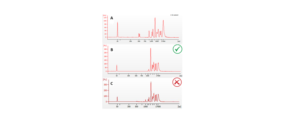 Figure 1. Size selection results of custom SPRI batch on 500 ng of a 1 kb extend ladder (NEB). A. unselected control. B. expected level of selection with removal of ≤1.kb marker. C. custom SPRI batch showing weak selection with retention of marker ≤1.5.
- Fragment size (FEMTO pulse):
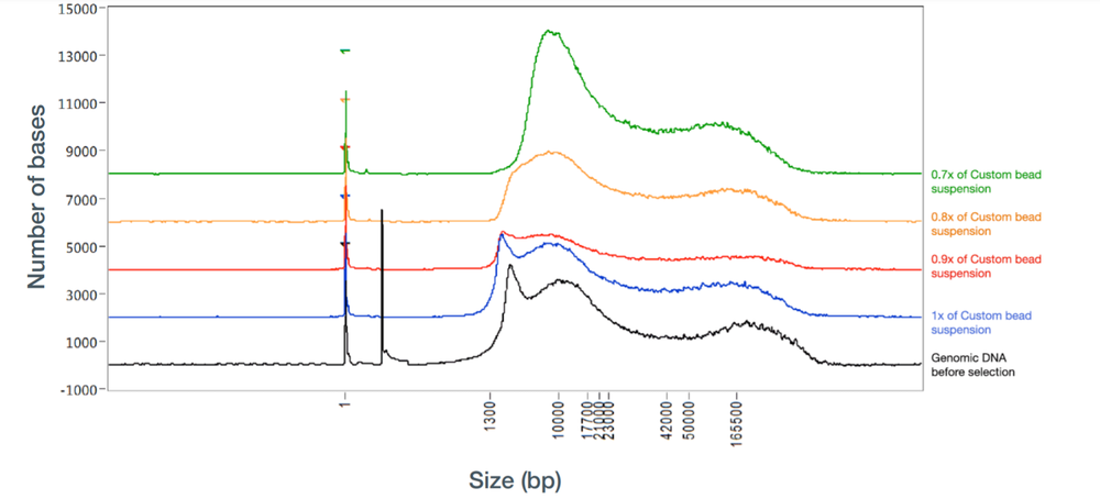 Figure 2. FEMTO pulse graph illustrating 0.7X beads (green line) provided the best selection of DNA >1.5-2 kb
Sequencing performance
Libraries for nanopore sequencing were prepared using the Ligation Sequencing Kit, with g-TUBE fragmentation. Read length profile after fragmentation and sequencing for gDNA samples which have, or have not had size selection performed:
Without size selection:
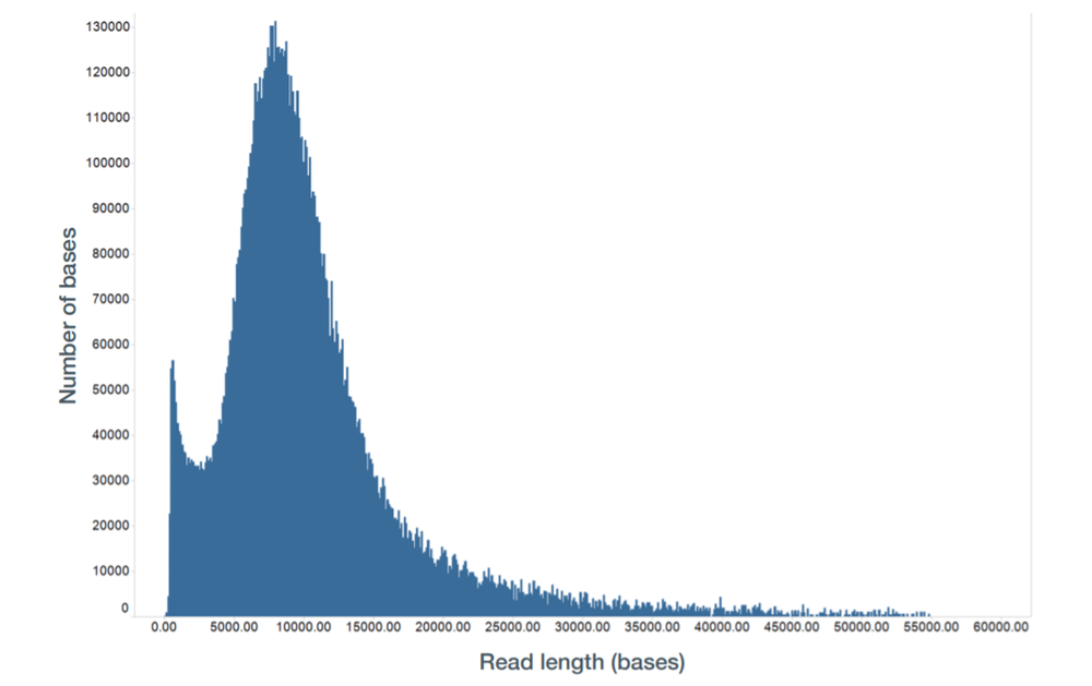 Figure 3. Read length of a DNA library prepared with the Ligation Sequencing Kit without size selection.
With size selection:
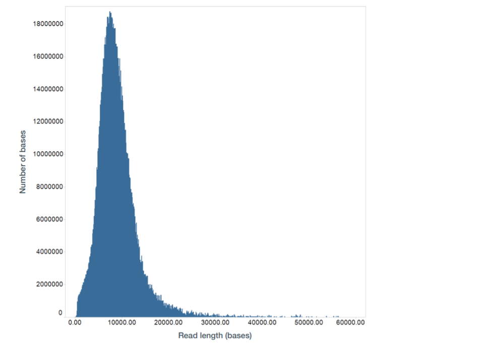 Figure 4. Read length of a DNA library prepared with the Ligation Sequencing Kit with size selection.
Changelog
| Version, date | Changelog |
|---|---|
| V3, 06th December 2024 | Formatting updates |
| V2, 26th September 2023 | Include additional optional QC step for batch validation and updated the customer buffer to use 40% w/v PEG 8000 rather than 50% to improve liquid handling. |
| V1, 28th January 2022 | Initial release |


