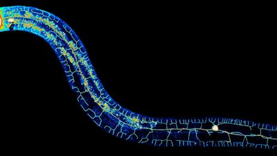
Temporary adhesion of the proseriate flatworm Minona ileanae
- Published on: September 9 2019
- Source: Philosophical Transactions of the Royal Society B
Flatworms can very rapidly attach to and detach from many substrates. In the presented work, we analysed the adhesive system of the marine proseriate flatworm Minona ileanae. We used light-, scanning- and transmission electron microscopy to analyse the morphology of the adhesive organs, which are located at the ventral side of the tail-plate. We performed transcriptome sequencing and differential RNA-seq for the identification of tail-specific transcripts. Using in situ hybridization expression screening, we identified nine transcripts that were expressed in the cells of the adhesive organs. Knock-down of five of these transcripts by RNA interference led to a reduction of the animal's attachment capacity. Adhesive proteins in footprints were confirmed using mass spectrometry and antibody staining. Additionally, lectin labelling of footprints revealed the presence of several sugar moieties. Furthermore, we determined a genome size of about 560 Mb for M. ileanae. We demonstrated the potential of Oxford Nanopore sequencing of genomic DNA as a cost-effective tool for identifying the number of repeats within an adhesive protein and for combining transcripts that were fragments of larger genes. A better understanding of the molecules involved in flatworm bioadhesion can pave the way towards developing innovative glues with reversible adhesive properties.


