Rapid metagenomic sequencing for surveillance of bacterial, fungal and viral pathogens using SQK-RPB114.24 (RMS_9215_v114_revF_18Nov2025)
ドキュメントオプション
MinION: Protocol
V RMS_9215_v114_revF_18Nov2025
FOR RESEARCH USE ONLY
Contents
Introduction to the protocol
Sample preparation
Library preparation
- 4. Reverse transcription and PCR
- 5. Clean-up, quantification and adapter attachment
- 6. Priming and loading the MinION and GridION Flow Cell
Sequencing and data analysis
Troubleshooting
1. Overview of protocol
This is an Early Access product.
For more information about our Early Access programmes, please see this article on product release phases.
Please ensure you always use the most recent version of the protocol.
Introduction to the rapid metagenomic sequencing protocol
This protocol outlines a method to perform agnostic metagenomic sequencing from extracted nucleic acid.
The method offers two options for sample preparation depending on your target sample and input:
- The DNA-only bacterial/fungal sample preparation utilises SQK-RPB114.24 reagents to tagment all DNA in the extract for amplification and sequencing.
- The DNA/RNA viral sample preparation has been optimised by Oxford Nanopore Technologies, and is derived from a method established by Josh Quick and Ingra M. Claro which utilises a shotgun approach using 9N primers to randomly reverse transcribe RNA and subsequently PCR-amplify DNA/RNA present in a sample.
Other areas of note:
- The performance of this method is reliant on sample type and was optimised for respiratory samples.
- While effort is made to reduce host background, sample types that are less complex/have less nucleic acid background are likely to perform better.
This protocol uses the Rapid PCR Barcoding Kit V14 (SQK-RPB114.24), which allows the potential to use up to 24 barcodes in one sequencing experiment.
For more information and example data from using this protocol please see our Rapid metagenomic sequencing for surveillance of bacterial, fungal and viral pathogens using SQK-RPB114.24 know-how document.
Steps in the sequencing workflow:
Prepare for your experiment
You will need to:
- Ensure you have your sequencing kit, the correct equipment and third-party reagents.
- Extract your DNA/RNA if not using the respiratory sample processing workflow in this protocol.
- Download the software for acquiring and analysing your data.
- Check your flow cell to ensure it has enough pores for a good sequencing run.
Host depletion and extraction
- If using the recommended respiratory sample host depletion and extraction, ensure you have the correct third-party reagents.
- Ensure you have fresh samples to use (not recommended for frozen/deactivated samples).
Library preparation
The Table below is an overview of the steps required in the library preparation, including timings and optional stopping points.
| Step | Process | Time | Stop option |
|---|---|---|---|
| Viral samples: reverse transcription | Reverse transcribe your RNA samples with the RLB RT 9N primer mix and the TSOmG template-switching oligo. | ~90 minutes | |
| Bacterial/fungal samples: DNA tagmentation | Tagment your DNA using the Fragmentation Mix from the sequencing kit. | 10 minutes | |
| PCR amplification | PCR your sample using the barcoded primer supplied in the sequencing kit. | 180 minutes | Optional: The PCR amplification can be performed and left at the hold temperature overnight. |
| Sample quantification and pooling | Quantify your barcoded PCR samples and pool them in equimolar ratios. | 15 minutes | |
| Clean-up and quantification | Perform a purification on your pooled samples and quantify. | 15 minutes | 4°C overnight |
| Rapid adapter attachment and clean-up | Attach the sequencing adapters to the DNA ends. | 5 minutes | We strongly recommend sequencing your library as soon as it is adapted. |
| Priming and loading the flow cell | Prime the flow cell and load the prepared library for sequencing. | 10 minutes |
Sequencing and analysis
You will need to:
- Start a sequencing run using the MinKNOW software, which will collect raw data from the device and convert it into basecalled reads.
- Analyse your data using the wf-metagenomics workflow available through EPI2ME or the command line.
Compatibility of this protocol
This protocol should only be used in combination with:
- Rapid PCR Barcoding Kit (SQK-RPB114.24)
- R10.4.1 flow cells (FLO-MIN114)
- Flow Cell Wash Kit (EXP-WSH004)
- Rapid Adapter Auxiliary V14 (EXP-RAA114)
- Sequencing Auxiliary Vials V14 (EXP-AUX003)
- Flow Cell Priming Kit V14 (EXP-FLP004)
- MinION Mk1D - MinION Mk1D IT requirements document
- GridION - GridION IT requirements document
2. Equipment and consumables
材料
- (FOR SAMPLE PREPARATION) Bacterial/fungal or viral/acellular sample inputs (see details below)
- (FOR LIBRARY PREPARATION) Extracted nucleic acid
- Rapid PCR Barcoding Kit 24 V14 (SQK-RPB114.24)
消耗品
- MinionとGridIONのFlow Cell
- Sputasol (Fisher, 11943262)
- RT-PCR Grade Water (Fisher, 10289774)
- Phosphate Buffer Saline (PBS) (Fisher, 11593377)
- 2X HL-SAN Buffer (4.5 M NaCl and 100 mM MgCl2) formulated using the two reagents below:
- • Invitrogen 5M NaCl (Fisher, 10255984)
- • Invitrogen 1M MgCl2 (Fisher, 10418464)
- HL-SAN Triton Free DNase (ArticZymes, 70911-202)
- Saponin solution (0.2% saponin in PBS) formulated using the reagent below:
- • Saponin (Sigma-Aldrich, 47036-50G-F)
- Matrix Lysing E tubes (Fisher, 11452420)
- MagMAX Viral/Pathogen Nucleic Acid Isolation Kit (Fisher, 16346582)
- 12 µM RLB RT 9N primer (IDT - Sequence: 5’-TTTTTCGTGCGCCGCTTCAACNNNNNNNNN-3’)
- 12 µM TSOmG template-switching oligo (IDT - Sequence: 5’-GCTAATCATTGCTTTTTCGTGCGCCGCTTCAACATmGmGmG-3’)
- 10 mM dNTPs (Fisher, 10610851)
- Maxima H Minus Reverse Transcriptase (Fisher, 13233159)
- RNaseOUT (Fisher, 10777019)
- LongAmp Hot Start Taq 2X Master Mix (NEB, M0533)
- Thermolabile Exonuclease I (NEB, M0568)
- Agencourt AMPure XP beads (Beckman Coulter™, A63881)
- Nuclease-free water (e.g. ThermoFisher, AM9937)
- Bovine Serum Albumin (BSA) (50 mg/ml) (e.g Invitrogen™ UltraPure™ BSA 50 mg/ml, AM2616)
- 1.5 ml Eppendorf DNA LoBind tubes
- 0.2 ml 薄壁のPCRチューブ
- Qubit dsDNA HS Assay Kit (ThermoFisher, Q32851)
- Qubit™ Assay Tubes (Invitrogen, Q32856)
装置
- MinIONかGridION のデバイス
- MinIONとGridIONのFlow Cell ライトシールド
- Benchtop microcentrifuge (max speed 15,000 rpm)
- 小型遠心機
- タイマー
- ボルテックスミキサー
- Thermomixer set at 37°C
- Thermomixer set at 65°C
- マグネットラック
- Bead-beater (e.g. FastPrep-24™ Classic bead beating grinder and lysis system)
- サーマルサイクラー
- 96-well PCR block
- アイスバケツ(氷入り)
- Hula mixer(緩やかに回転するミキサー)
- Qubit fluorometer (or equivalent)
- P1000 ピペット及びチップ
- P200 ピペットとチップ
- P100 ピペットとチップ
- P20 ピペットとチップ
- P10 ピペットとチップ
- P2 ピペットとチップ
オプション装置
- Suitable biosafety equipment for your sample(s) (e.g. Microbiological Safety Cabinet II contamination level 3 or equivalent)
Ensure pathogenic samples are handled in appropriate biosafety conditions.
Please adhere to the correct health and safety practices in accordance to your laboratory standards and local rules and regulations.
Note: We recommend as a minimum the use of a Class II Biological Safety Cabinet. Please consider that some organisms can survive bead-beating steps during extraction. Ensure you are taking the necessary precautions during your sample and library preparation.
For this protocol you will need the following sample input(s):
For sample preparation:
- ≥ 250 µl of Sputum/Endotracheal aspirates
OR
- ≥ 250 µl of Bronchoalveolar lavages/Mini-BALs
OR
- ≥ 500 µl of VTM/UTM-stored oral/nasal swabs (only suitable for viral samples)
Note: If quantities allow, a sample can be processed for both bacterial/fungal sample preparation and viral sample preparation. Please note, each preparation type will differ in method and should be treated as a separate sample.
For library preparation:
- Extracted nucleic acid from the sample types above.
Input DNA/RNA
How to QC your input
It is important that the input DNA/RNA meets the quantity and quality requirements. Using too little or too much DNA/RNA, or DNA/RNA of poor quality (e.g. highly fragmented or containing RNA or chemical contaminants) can affect your library preparation.
For instructions on how to perform quality control of your DNA/RNA sample, please read the Input DNA/RNA QC protocol.
Chemical contaminants
Depending on how the DNA/RNA is extracted from the raw sample, certain chemical contaminants may remain in the purified DNA/RNA, which can affect library preparation efficiency and sequencing quality. Read more about contaminants on the Contaminants page of the Community.
Input controls
Negative internal controls
We recommend a negative control is included and processed as a real sample through the entire process to monitor for contamination events.
The negative control can be PBS or a third-party negative sample matrix.
Third-party reagents
We have validated and recommend the use of all the third-party reagents used in this protocol. Alternatives have not been tested by Oxford Nanopore Technologies.
For all third-party reagents, we recommend following the manufacturer's instructions to prepare the reagents for use.
Agencourt AMPure XP beads
Additional Agencourt AMPure XP beads may be required alongside the AMPure XP Beads (AXP) provided in the sequencing kit for the clean-up step following PCR amplification.
Eppendorf tube orientation in centrifuge
For all centrifugation steps, ensure that tubes are loaded into the centrifuge with the hinge side of the tube facing outwards. This will assist in visual identification of the pellet.
Ensure gentle handling when removing the tubes from the centrifuge to avoid dislodging the pellet.

Check your flow cell
We highly recommend that you check the number of pores in your flow cell prior to starting a sequencing experiment. This should be done within 12 weeks of purchasing your MinION/GridION Flow Cells. Oxford Nanopore Technologies will replace any unused flow cell with fewer than the number of pores listed in the Table below, when the result is reported within two days of performing the flow cell check, and when the storage recommendations have been followed. To do the flow cell check, please follow the instructions in the Flow Cell Check document.
| Flow cell | Minimum number of active pores covered by warranty |
|---|---|
| MinION/GridION Flow Cell | 800 |
The Rapid Adapter (RA) used in this kit and protocol is not interchangeable with other sequencing adapters.
Rapid PCR Barcoding Kit 24 V14 (SQK-RPB114.24) contents
We are in the process of reformatting the Rapid Barcode Primers provided in this kit into a plate format. Additionally we have lowered the concentration of the Rapid Barcode Primers in the plate format to 1 µM (down from 10 µM in the vial format). These changes will reduce plastic waste and facilitates automated applications.
Please ensure you follow the correct instructions in the method for your kit format and Rapid Barcode Primers concentration.
Plate format with Rapid Barcode Primers at 1 µM
| Name | Acronym | Cap colour | No. of vials | Fill volume per vial (µl) |
|---|---|---|---|---|
| Fragmentation Mix | FRM | 1 | Brown | 160 |
| Rapid Adapter | RA | 1 | Green | 15 |
| Adapter Buffer | ADB | 1 | Clear | 100 |
| AMPure XP Beads | AXP | 3 | Amber | 1,200 |
| Elution Buffer | EB | 2 | Black | 500 |
| EDTA | EDTA | 1 | Blue | 700 |
| Sequencing Buffer | SB | 1 | Red | 700 |
| Library Beads | LIB | 1 | Pink | 600 |
| Library Solution | LIS | 1 | White cap, pink label | 600 |
| Flow Cell Flush | FCF | 1 | Clear cap, light blue label | 8,000 |
| Flow Cell Tether | FCT | 1 | Purple | 200 |
| Rapid Barcode Primers (01-24) at 1 µM | RPB | - | 2 plates, 3 sets of primer barcodes per plate | 15 µl per well |
Note: This product contains AMPure XP reagent manufactured by Beckman Coulter, Inc. and can be stored at -20°C with the kit without detriment to reagent stability.
Vial format with Rapid Barcode Primers at 10 µM
| Name | Acronym | Cap colour | No. of vials | Fill volume per vial (µl) |
|---|---|---|---|---|
| Fragmentation Mix | FRM | Brown | 1 | 160 |
| Rapid Adapter | RA | Green | 1 | 15 |
| Adapter Buffer | ADB | Clear | 1 | 100 |
| AMPure XP Beads | AXP | Amber | 3 | 1,200 |
| Elution Buffer | EB | Black | 2 | 500 |
| EDTA | EDTA | Blue | 1 | 700 |
| Sequencing Buffer | SB | Red | 1 | 700 |
| Library Beads | LIB | Pink | 1 | 600 |
| Library Solution | LIS | White cap, pink label | 1 | 600 |
| Flow Cell Flush | FCF | Clear | 1 | 8,000 |
| Flow Cell Tether | FCT | Purple | 1 | 200 |
| Rapid Barcode Primer 01-24 | RLB01-24 | Clear | 24 (one per barcode) | 15 |
Note: This product contains AMPure XP reagent manufactured by Beckman Coulter, Inc. and can be stored at -20°C with the kit without detriment to reagent stability.
Rapid Barcode Primers
| Component | Sequence |
|---|---|
| Rapid Barcode Primer 01 | AAGAAAGTTGTCGGTGTCTTTGTG |
| Rapid Barcode Primer 02 | TCGATTCCGTTTGTAGTCGTCTGT |
| Rapid Barcode Primer 03 | GAGTCTTGTGTCCCAGTTACCAGG |
| Rapid Barcode Primer 04 | TTCGGATTCTATCGTGTTTCCCTA |
| Rapid Barcode Primer 05 | CTTGTCCAGGGTTTGTGTAACCTT |
| Rapid Barcode Primer 06 | TTCTCGCAAAGGCAGAAAGTAGTC |
| Rapid Barcode Primer 07 | GTGTTACCGTGGGAATGAATCCTT |
| Rapid Barcode Primer 08 | TTCAGGGAACAAACCAAGTTACGT |
| Rapid Barcode Primer 09 | AACTAGGCACAGCGAGTCTTGGTT |
| Rapid Barcode Primer 10 | AAGCGTTGAAACCTTTGTCCTCTC |
| Rapid Barcode Primer 11 | GTTTCATCTATCGGAGGGAATGGA |
| Rapid Barcode Primer 12 | GTTGAGTTACAAAGCACCGATCAG |
| Rapid Barcode Primer 13 | AGAACGACTTCCATACTCGTGTGA |
| Rapid Barcode Primer 14 | AACGAGTCTCTTGGGACCCATAGA |
| Rapid Barcode Primer 15 | AGGTCTACCTCGCTAACACCACTG |
| Rapid Barcode Primer 16 | CGTCAACTGACAGTGGTTCGTACT |
| Rapid Barcode Primer 17 | ACCCTCCAGGAAAGTACCTCTGAT |
| Rapid Barcode Primer 18 | CCAAACCCAACAACCTAGATAGGC |
| Rapid Barcode Primer 19 | GTTCCTCGTGCAGTGTCAAGAGAT |
| Rapid Barcode Primer 20 | TTGCGTCCTGTTACGAGAACTCAT |
| Rapid Barcode Primer 21 | GAGCCTCTCATTGTCCGTTCTCTA |
| Rapid Barcode Primer 22 | ACCACTGCCATGTATCAAAGTACG |
| Rapid Barcode Primer 23 | CTTACTACCCAGTGAACCTCCTCG |
| Rapid Barcode Primer 24 | GCATAGTTCTGCATGATGGGTTAG |
3. Host depletion and extraction
材料
- Sputum/Endotracheal aspirates
- Bronchoalveolar lavages/Mini-BALs
- VTM/UTM-stored oral/nasal swabs (viral-arm only)
消耗品
- Sputasol (Fisher, 11943262)
- RT-PCR Grade Water (Fisher, 10289774)
- Phosphate Buffer Saline (PBS) (Fisher, 11593377)
- 2X HL-SAN Buffer (4.5 M NaCl and 100 mM MgCl2) formulated using the two reagents below:
- • Invitrogen 5M NaCl (Fisher, 10255984)
- • Invitrogen 1M MgCl2 (Fisher, 10418464)
- HL-SAN Triton Free DNase (ArticZymes, 70911-202)
- Saponin solution (0.2% saponin in PBS) formulated using the reagent below:
- • Saponin (Sigma-Aldrich, 47036-50G-F)
- Matrix Lysing E tubes (Fisher, 11452420)
- MagMAX Viral/Pathogen Nucleic Acid Isolation Kit (Fisher, 16346582)
- 1.5 ml Eppendorf DNA LoBind tubes
- 15 ml Falcon tubes
装置
- Benchtop microcentrifuge (max speed 15,000 rpm)
- 小型遠心機
- タイマー
- ボルテックスミキサー
- Thermomixer set at 37°C
- Thermomixer set at 65°C
- マグネットラック
- Bead-beater (e.g. FastPrep-24™ Classic bead beating grinder and lysis system)
- P1000 ピペット及びチップ
- P200 ピペットとチップ
- P20 ピペットとチップ
- P10 ピペットとチップ
オプション装置
- Suitable biosafety equipment for your sample(s) (e.g. Microbiological Safety Cabinet II contamination level 3 or equivalent)
Ensure you are keeping your bacterial/fungal samples separate from your viral samples.
If processing a mix of bacterial/fungal samples and viral samples, ensure you keep them separate to avoid following the wrong sample preparation method.
Both methods can be followed simultaneously if processing different sample types prior to library preparation. Please ensure you are following the correct method for each sample type.
Note: A sample can be processed for both bacterial/fungal sample preparation and viral sample preparation. Please follow the instructions below, where the supernatant is used for viral sample preparation and the pellet is used for bacterial/fungal sample preparation. Please note, each preparation type will differ in method and should be treated as a separate sample.
Ensure you have sufficient sample input. You will need:
- 250 µl of sputum, endotracheal aspirate or other viscous samples (e.g. mucosal BAL).
OR
- 500 µl of swab sample in transport media or other liquid samples.
Input controls
Negative internal controls
We recommend a negative control is included and processed as a real sample through the entire process to monitor for contamination events.
The negative control can be PBS or a third-party negative sample matrix.
Prepare a working solution of Sputasol in nuclease-free water (1:13.33) as follows:
- In a fresh 15 ml Falcon tube, dispense 750 µl Sputasol.
- Add 9.25 ml nuclease-free water.
- Throughly mix by vortexing.
Note: Volumes can be adjusted to meet your experiment requirements.
Prepare a 0.2% saponin-PBS solution:
| Reagent | Quantity/Volume |
|---|---|
| Saponin (Sigma-Aldrich, 47036-50G-F) | 0.01 g |
| Phosphate Buffer Saline (PBS) | 5,000 µl |
| Total volume 0.2% saponin-PBS solution | 5,000 µl |
Note: Quantity/volumes can be adjusted to meet your experiment requirements.
The 0.2% saponin-PBS solution can be stored in the fridge (~4°C) for up to 30 days.
Prepare the 2X HLSAN Buffer (4.5 M NaCl and 100 mM MgCl2) as follows:
| Reagent | Volume |
|---|---|
| Invitrogen 5M NaCl | 4,500 µl |
| Invitrogen 1M MgCl2 | 500 µl |
| Total | 5,000 µl |
Note: Volumes can be adjusted to meet your experiment requirements.
The formulated 2X HLSAN Buffer can be stored at room temperature (~22°C) long-term.
Ensure your thermomixers are set to 37°C and 65°C.
Prepare your sputum, endotracheal (ETT), or any other mucoid sample(s):
Note: Transport Media and BAL samples do not require this unless they are mucoid.
- To each sample, add an equal volume (1:1) of Sputasol working solution.
- Mix by vortexing for 30 seconds.
- Incubate at room temperature until liquefication (at least 5 minutes).
Tip: If the sample is still viscous or sticky after 5 minutes, repeat the process above. Full liquefication of sputum is important for good depletion and efficient extraction.
For each sample, transfer 500 µl into a separate clean 1.5 ml Eppendorf tube.
Centrifuge for 5 minutes at 10,000 x g.
Carefully remove tubes from centrifuge without disturbing the solution.
Remember, your sample(s) can be processed for both viral sample preparation and bacterial/fungal sample preparation.
Please follow the instructions outlined below and treat each preparation as a separate sample.
Perform sample preparation side-by-side depending on your sample type:
| Step | For viral sample preparation | For bacterial/fungal samples |
|---|---|---|
| 1. | For each sample, transfer 300 µl of the supernatant (by aspirating from the top) to a separate new 1.5 ml Eppendorf tube. | OPTIONAL: the remaining pellet and supernatant from the sample used in the viral sample preparation can be taken forward and processed in the bacterial/fungal sample preparation as seen below |
| 2. | Using a pipette, carefully remove most of the supernatant without disturbing the pellet, leaving enough volume to cover the pellet. Note: Ensure there is enough supernatant left (~50 µl) above the pellet so as to not disturb it. | |
| 3. | Thoroughly resuspend the pellet in 200 µl of the prepared 0.2% saponin-PBS solution. Mix by pipetting. | |
| 4. | Add the depletion reagent to the sample supernatant: • Add 10 µl of HLSAN enzyme. • Vortex mix for 3 seconds. | Add the depletion reagents to the resuspended pellet as follows: • Add 200 µl of the prepared 2X HLSAN Buffer. • Add 10 µl of HLSAN enzyme. • Vortex mix for 3 seconds. |
| 5. | Incubate the reaction on a thermomixer at 37°C for 10 minutes at 1,000 rpm. | Incubate the reaction on a thermomixer at 37°C for 10 minutes at 1,000 rpm. |
| 6. | To each tube containing the sample supernatant, prepare the following reaction: 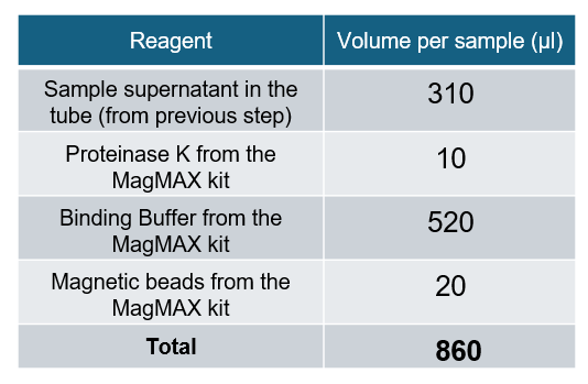 Ensure the reagents are mixed by inverting the tube multiple times. Ensure the reagents are mixed by inverting the tube multiple times. Avoid mixing the lysis reagents too vigorously or with any method that would lead to foam (e.g. vortexing). Note: A mastermix of the extraction reagents can be made and added together instead. | |
| 7. | Add 900 µl of PBS to your sample(s) and mix by pipetting. | |
| 8. | Centrifuge your sample(s) at 12,000 x g for 3 minutes. | |
| 9. | Using a pipette, carefully remove most of the supernatant without disturbing the pellet, leaving enough volume to cover the pellet. Ensure there is enough supernatant left (~50 µl) above the pellet so as to not disturb it. | |
| 10. | Resuspend pellet in 500 µl of PBS and mix by pipetting. | |
| 11. | For each sample, transfer whole volume of sample into a separate new bead-beating tube (matrix lysing E tubes). | |
| 12. | Insert the bead-beating tubes into the FastPrep-24 Classic bead beating grinder and lysis system and run the program as follows:  | |
| 13. | Remove the bead-beating tubes from the Fast-prep device, and centrifuge for 30 seconds at max speed (>20,000 x g). | |
| 14. | For each sample, transfer 300 µl of the supernatant to a separate new 1.5 ml Eppendorf tube. Note: Avoid aspirating the beads from the bead-beating tube. | |
| 15. | To each tube containing the sample supernatant, prepare the following reaction: 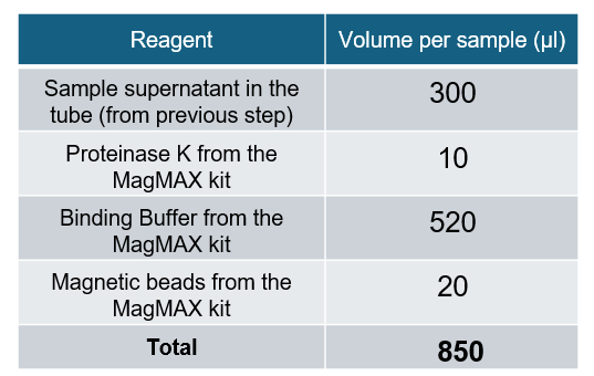 Ensure the reagents are mixed by inverting the tube multiple times. Ensure the reagents are mixed by inverting the tube multiple times. Avoid mixing the lysis reagents too vigorously or with any method that would lead to foam (e.g. vortexing). Note: A mastermix of the extraction reagents can be made and added together instead. |
Transfer all sample tubes to a thermomixer and incubate the reactions at 65°C for 5 minutes at 1,000 rpm.
Transfer the sample tube(s) to a hulamixer and incubate/mix at room temperature for 5 min.
Hulamixer settings:
- Orbital 15 rpm (10 seconds)
- Reciprocal 70° (15 seconds)
- Vibro/pause 5° (3 seconds)
- 5 minutes total
Note: Tube inversion is crucial during this incubation, as the beads may collect at the bottom of the tube. We highly recommend the use of a hulamixer, however if this is not available, ensure the samples are manually inverted during the incubation.
Prepare 2 ml of fresh 80% ethanol per sample in nuclease-free water.
Briefly spin down the tube(s) and pellet on a magnetic rack until supernatant is clear and colourless (for at least 5 minutes). Keep the tube on the magnetic rack, and pipette off the supernatant.
Take care not to disturb the pelleted beads.
Remove the tube from the magnetic rack and add 1 ml of Wash Buffer from the MagMAX kit. Gently mix by inverting the tube until fully resuspended.
Note: Avoid techniques that will create foam when mixing your tubes. This can negatively impact sample recovery.
Briefly spin down the tube(s) and pellet on a magnetic rack until supernatant is clear and colourless (for at least 2 minutes). Keep the tube on the magnetic rack, and pipette off the supernatant.
Remove the tube from the magnetic rack and add 1 ml of 80% ethanol. Gently mix by inverting the tube until fully resuspended.
Briefly spin down the tube(s) and pellet on a magnetic rack until supernatant is clear and colourless (for at least 2 minutes). Keep the tube on the magnetic rack, and pipette off the supernatant.
Remove the tube from the magnetic rack and add 500 µl of 80% ethanol. Gently mix by inverting the tube until fully resuspended.
Briefly spin down the tube(s) and pellet on a magnetic rack until supernatant is clear and colourless (for at least 2 minutes). Keep the tube on the magnetic rack, and pipette off the supernatant.
Keeping the tube on the magnetic rack, leave the lid open and allow to dry for ~2 minutes, but do not dry the pellet to the point of cracking.
Remove the tube from the magnetic rack and resuspend the pellet by pipetting in 20 µl of nuclease-free water. Ensure the pellet is fully resuspended by pipette mixing.
To aid with sample elution, transfer tubes to a thermomixer and incubate at 65°C for 5 minutes at 1,000 rpm.
Pellet the beads on a magnet until the eluate is clear and colourless for at least 1 minute.
Remove and retain the eluate (~15 µl) into a clean 1.5 ml Eppendorf DNA LoBind tube.
- Dispose of the pelleted beads
Take your extracted sample forward into the library preparation section of this protocol.
Alternatively, if you are not using your sample immediately, store at -20°C.
4. Reverse transcription and PCR
材料
- For viral sample preparation: 10 µl of extracted nucleic acid from previous step
- For bacterial/fungal sample preparation: 3 µl of extracted nucleic acid from previous step
- Fragmentation Mix (FRM)
- Rapid Barcode Primers (vial format (RLB01-24, at 10 µM) or plate format (RPB01-24, at 1 µM))
消耗品
- 12 µM RLB RT 9N primer (IDT - Sequence: 5’-TTTTTCGTGCGCCGCTTCAACNNNNNNNNN-3’)
- 12 µM TSOmG template-switching oligo (IDT - Sequence: 5’-GCTAATCATTGCTTTTTCGTGCGCCGCTTCAACATmGmGmG-3’)
- 10 mM dNTPs (Fisher, 10610851)
- Maxima H Minus Reverse Transcriptase (Fisher, 13233159)
- RNaseOUT (Fisher, 10777019)
- LongAmp Hot Start Taq 2X Master Mix (NEB, M0533)
- Nuclease-free water (e.g. ThermoFisher, AM9937)
- 1.5 ml Eppendorf DNA LoBind tubes
- 0.2 ml 薄壁のPCRチューブ
装置
- Hula mixer(緩やかに回転するミキサー)
- 小型遠心機
- サーマルサイクラー
- 96-well PCR block
- タイマー
- アイスバケツ(氷入り)
- P1000 ピペット及びチップ
- P200 ピペットとチップ
- P100 ピペットとチップ
- P20 ピペットとチップ
- P10 ピペットとチップ
- P2 ピペットとチップ
Ensure you are following the correct steps in the method for your sample type.
The method differs for bacterial/fungal sample processing and for viral sample processing. Please ensure you keep track of your samples to ensure you are following the correct method.
Note: If a sample has been processed for both bacterial/fungal sample preparation and viral sample preparation, you will have two "separate" samples to process and barcode in library preparation.
Thaw kit components at room temperature, then spin down briefly using a microfuge and mix as indicated by the Table below. Then place on ice:
| Reagents | 1. Thaw at room temperature | 2. Briefly spin down | 3. Mix well by pipetting |
|---|---|---|---|
| Fragmentation Mix (FRM) | Not frozen | ✓ | ✓ |
| Rapid Barcode Primers (vial format (RLB01-24, at 10 µM) or plate format (RPB01-24, at 1 µM)) | ✓ | ✓ | ✓ |
| RLB RT 9N primer (12 µM) | ✓ | ✓ | ✓ |
| TSOmG template-switching oligo (12 μM) | ✓ | ✓ | ✓ |
| 10 mM dNTP solution | ✓ | ✓ | ✓ |
| Maxima H Minus Reverse Transcriptase | Not frozen | ✓ | ✓ |
| Maxima H Minus 5x RT Buffer | ✓ | ✓ | ✓ |
| LongAmp Taq 2X Master Mix | ✓ | ✓ | ✓ |
| RNaseOUT | Not frozen | ✓ | ✓ |
The wells of the barcoding plate are intended for single use only. Please ensure your barcode well is sealed before use, and do not reuse the barcode well once pierced/opened.
Prepare each of your extracted nucleic acid samples from the previous step in a separate clean 0.2 ml tube:
For viral sample preparation:
- Take forward 10 µl of each viral nucleic acid extract.
Note: Quantifying viral samples is not recommended, as these will likely be below the limit of detection for Qubit High Sensitivity or similar fluorescence-based quantification methods. Please proceed with the recommended volume of viral nucleic acid extract.
For bacterial/fungal samples:
- Quantify 1 µl of each bacterial/fungal extraction using Qubit High Sensitivity.
- If the extract is more than 1 ng/µl: dilute to 1 ng/µl using nuclease-free water.
- If the extract is less than 1 ng/µl (or unquantifiable): no dilution is necessary, and extract is used directly.
- Take forward 3 µl of each bacterial/fungal DNA extract.
Perform the RT reaction / tagmentation reaction side-by-side depending on your sample type:
Note: If processing both sample types we recommend preparing the viral samples first as they have a longer incubation time. The bacterial/fungal samples can be processed during the viral sample incubation.
| Step | For viral sample preparation (RT reaction) | For bacterial/fungal samples (tagmentation) |
|---|---|---|
| 1. | In a clean 0.2 ml thin-walled PCR tube, prepare the following master mix. Tip: Generate sufficient volume for each viral sample preparation + 1 extra volume for excess: 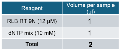 | |
| 2. | Mix the master mix prepared above by vortexing and spin down briefly. | |
| 3. | Add 2 µl of the prepared mastermix to each of your 10 µl of viral nucleic acid extract. | |
| 4. | Incubate the reaction at 65°C for 5 minutes, then transfer the samples to ice immediately. | |
| 5. | Keep your samples on ice for 2 minutes. | |
| 6. | In a clean 0.2 ml thin-walled PCR tube, prepare the Maxima H master mix. Tip: Generate sufficient volume for each viral sample preparation + 1 extra volume for excess: 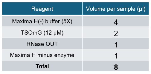 Note: Add the Maxima H Enzyme last. Keep the master mix on ice. Note: Add the Maxima H Enzyme last. Keep the master mix on ice. The master mix should be aliquoted to your samples and incubated relatively quickly, as the enzymes are not hot-start, and the reaction will start as soon as it comes in contact with your sample. | |
| 7. | Add 8 µl of the Maxima H master mix to each of your viral samples for a total combined volume of 20 µl. | |
| 8. | Pre-heat a PCR block to 42°C, then add your sample tubes and incubate using the following conditions: 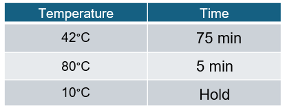 | During the RT reaction (viral samples), the bacterial/fungal samples can be prepared. |
| 9. | To each of the 3 µl of bacterial/fungal DNA extract, add 1 µl of FRM, and mix gently by flicking the tube. | |
| 10. | Pre-heat a PCR block to 30°C, then add your sample tubes and incubate using the following conditions: 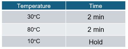 |
Please ensure you are following the correct method for your SQK-RPB114.24 kit format.
In addition to the kit format change, we have lowered the concentration of the Rapid Barcode Primers in the plate format to 1 µM (down from 10 µM in the vial format) to facilitate automation usage and improve kit usability.
Please note, this will mean that the volumes used in the PCR reaction set up will vary depending on the kit format you have available.
In a clean 1.5 Eppendorf tube, prepare the following master mix:
Tip: Generate sufficient volume for each sample preparation + 1 extra volume for excess:
Plate format with Rapid Barcode Primers at 1 µM
| Reagent | Volume |
|---|---|
| LongAmp Hot Start Taq 2X Master Mix | 25 µl |
| Nuclease-free water | 11 µl |
| Total volume | 36 µl |
Mix by pipetting, taking care not to generate bubbles or foam.
Vial format with Rapid Barcode Primers at 10 µM
| Reagent | Volume |
|---|---|
| LongAmp Hot Start Taq 2X Master Mix | 25 µl |
| Nuclease-free water | 20 µl |
| Total volume | 45 µl |
Mix by pipetting, taking care not to generate bubbles or foam.
Note: The non-hot start product, LongAmp Taq 2X Master Mix (e.g. NEB, M0287) is also compatible with this method. If using this alternative, please ensure you follow the manufacturers instructions and prepare this reaction on ice.
Aliquot out the master mix into a new separate 0.2 ml thin-walled PCR tube for each sample preparation.
To each tube containing the prepared master mix, add the Rapid Barcode Primers (01-24). Each tube should have a different barcode:
Note: To make sample tracking easier, we recommend that the two types of sample preparation are given different blocks of barcodes:
e.g. Barcodes 1-4 for the viral samples and barcodes 5-8 for the bacterial/fungal samples.
For plate format with Rapid Barcode Primers at 1 µM
| Reagent | Volume |
|---|---|
| Tube containing the prepared master mix (from previous step) | 36 µl |
| Rapid Barcode Primers (01-24, at 1 µM) | 10 µl |
| Total | 46 µl |
For vial format with Rapid Barcode Primers at 10 µM
| Reagent | Volume |
|---|---|
| Tube containing the prepared master mix (from previous step) | 45 µl |
| Rapid Barcode Primers (01-24, at 10 µM) | 1 µl |
| Total | 46 µl |
To each tube containing master mix and a Rapid Barcode Primer, add your RT (viral) sample or tagmented (bacterial/fungal) sample:
For viral samples:
- Add 5 µl RT sample to the reaction tube. Do not deviate from the recommended volume as this can lead to sub-optimal results.
| Reagent | Volume |
|---|---|
| Master mix + Rapid Barcode Primer | 46 µl |
| RT viral sample | 5 µl |
| Total | 51 µl |
Note: If you are concerned that you will not reach the minimum molarity, we recommend assembling two separate reactions for your viral sample using the volumes above, and pooling following PCR. Do not alter the PCR volumes as this will result in poor performance of the PCR and lead to sub-optimal results.
For bacterial/fungal samples:
- Add the full volume of tagmented sample to the reaction tube:
| Reagent | Volume |
|---|---|
| Master mix + Rapid Barcode Primer | 46 µl |
| Tagmented bacterial/fungal DNA sample | ~4 µl |
| Total | 50 µl |
Mix gently by flicking the tube, and spin down.
Amplify using the following cycling conditions:
| Cycle step | Temperature | Time | No. of cycles |
|---|---|---|---|
| Initial denaturation | 95°C | 45 seconds | 1 |
| Denaturation Annealing Extension | 95°C 56°C 65°C | 15 seconds 15 seconds 4 minutes | 30 |
| Final extension | 65°C | 6 minutes | 1 |
| Hold | 10°C | ∞ |
Note: Total PCR time is 2 hours 43 minutes. Timing may differ depending on equipment and ambient conditions.
Optional: The PCR amplification can be performed and left at the hold temperature overnight.
Take forward your sample into the clean up, quantification and adapter attachment step.
5. Clean-up, quantification and adapter attachment
材料
- AMPure XP Beads (AXP)
- Elution Buffer (EB)
- Rapid Adapter (RA)
- Adapter Buffer (ADB)
消耗品
- Thermolabile Exonuclease I (NEB, M0568)
- nuclease-free waterで調整した 80% エタノール溶液
- 1.5 ml Eppendorf DNA LoBind tubes
- 0.2 ml 薄壁のPCRチューブ
- Qubit dsDNA HS Assay Kit (ThermoFisher, Q32851)
- Qubit™ Assay Tubes (Invitrogen, Q32856)
装置
- サーマルサイクラー
- Hula mixer(緩やかに回転するミキサー)
- マグネットラック
- Qubit fluorometer (or equivalent)
- タイマー
- P1000 ピペット及びチップ
- P200 ピペットとチップ
- P100 ピペットとチップ
- P20 ピペットとチップ
- P10 ピペットとチップ
- P2 ピペットとチップ
Check your flow cell.
We recommend performing a flow cell check before completing your library prep to ensure you have a flow cell with enough pores for a good sequencing run.
See the flow cell check document for more information.
Quantify the sample tubes (PCR products from the previous step) using the Qubit dsDNA HS Assay Kit.
Make a note of each samples concentration.
Note: Sample concentration may vary depending on input. Some of your samples might be below the limit of detection for the Qubit dsDNA HS Assay Kit. This is not an uncommon occurance.
Please proceed with all of your samples, following the recommendations in the instructions below.
Add 1 µl of Thermolabile Exo I to each of the sample tubes (PCR product) and mix by pipetting.
Incubate the reactions using the following conditions:
| Temperature | Time |
|---|---|
| 37°C | 10 minutes |
| 80°C | 1 minute |
| 10°C | Hold |
In a new 1.5 ml Eppendorf DNA LoBind tube, pool all barcoded samples in equimolar ratios to a combined final concentration of 800 ng.
For example, if 10 barcodes were used, take forward 80 ng of each sample.
Note: In cases where the concentration of your sample PCR product is too low, take forward the maximum available volume.
Resuspend the AMPure XP beads (AXP) by vortexing.
Add 0.6X volume of AMPure XP beads (AXP) to the pooled samples.
Ensure you have accurately measured the volume of your pooled samples to maintain the 0.6X ratio of the AMPure XP beads (AXP).
Incubate on a Hula mixer (rotator mixer) for 5 minutes at room temperature.
Prepare at least 1 ml of fresh 80% ethanol in nuclease-free water.
Briefly spin down the sample and pellet on a magnetic rack until supernatant is clear and colourless. Keep the tube on the magnetic rack, and pipette off the supernatant.
Keep the tube on the magnet and wash the beads with 500 µl of freshly-prepared 80% ethanol without disturbing the pellet. Remove the ethanol using a pipette and discard.
Repeat the previous step.
Spin down and place the tube back on the magnet. Pipette off any residual ethanol. Allow to dry for ~30 seconds, but do not dry the pellet to the point of cracking.
Remove the tube from the magnetic rack and resuspend the pellet in 15 µl Elution Buffer (EB).
Incubate for 2 minutes at room temperature.
Pellet the beads on a magnet until the eluate is clear and colourless.
Remove and retain 14 µl of eluate containing the DNA library into a clean 1.5 ml Eppendorf DNA LoBind tube.
- Remove and retain the eluate which contains the DNA library in a clean 1.5 ml Eppendorf DNA LoBind tube.
- Dispose of the pelleted beads.
Quantify 1 µl of eluted sample using a Qubit fluorometer and the Qubit dsDNA HS Assay Kit.
Expected yield of 10-40 ng/µl
Take forward 11 µl of your eluted samples into a clean 1.5 ml Eppendorf DNA LoBind tube.
In a fresh 1.5 ml Eppendorf DNA LoBind tube, dilute the Rapid Adapter (RA) as follows and pipette mix:
| Reagent | Volume |
|---|---|
| Rapid Adapter (RA) | 1.5 μl |
| Adapter Buffer (ADB) | 3.5 μl |
| Total | 5 μl |
Add 1 µl of the diluted Rapid Adapter (RA) to the barcoded DNA.
Mix gently by flicking the tube, and spin down.
Incubate the reaction for 5 minutes at room temperature.
The prepared library is used for loading into the flow cell. Store the library on ice or at 4°C until ready to load.
6. Priming and loading the MinION and GridION Flow Cell
材料
- Flow Cell Flush (FCF)
- Flow Cell Tether (FCT)
- Library Beads (LIB)
- Sequencing Buffer (SB)
消耗品
- MinionとGridIONのFlow Cell
- Bovine Serum Albumin (BSA) (50 mg/ml) (e.g Invitrogen™ UltraPure™ BSA 50 mg/ml, AM2616)
- 1.5 ml Eppendorf DNA LoBind tubes
装置
- MinIONかGridION のデバイス
- MinIONとGridIONのFlow Cell ライトシールド
- P1000 ピペット及びチップ
- P100 ピペットとチップ
- P20 ピペットとチップ
- P10 ピペットとチップ
Please note, this kit is only compatible with R10.4.1 flow cells (FLO-MIN114).
Take the flow cell out of the fridge and leave it at room temperature for 20 minutes. This will improve visibility of the array during priming and sample loading.
Priming and loading a flow cell
We recommend all new users watch the 'Priming and loading your flow cell' video before your first run.
Thaw the Sequencing Buffer (SB), Library Beads (LIB), Flow Cell Tether (FCT) and Flow Cell Flush (FCF) at room temperature before mixing by vortexing. Then spin down and store on ice.
For optimal sequencing performance and improved output on MinION R10.4.1 flow cells (FLO-MIN114), add Bovine Serum Albumin (BSA) to the flow cell priming mix at a final concentration of 0.2 mg/ml.
Note: We do not recommend using any other albumin type (e.g. recombinant human serum albumin).
To prepare the flow cell priming mix with BSA, combine the following reagents in a fresh 1.5 ml Eppendorf DNA LoBind tube. Mix by inverting the tube and pipette mix at room temperature:
| Reagents | Volume per flow cell |
|---|---|
| Flow Cell Flush (FCF) | 1,170 µl |
| Bovine Serum Albumin (BSA) at 50 mg/ml | 5 µl |
| Flow Cell Tether (FCT) | 30 µl |
| Final total volume in tube | 1,205 µl |
Open the MinION or GridION device lid and slide the flow cell under the clip. Press down firmly on the priming port cover to ensure correct thermal and electrical contact.
Complete a flow cell check to assess the number of pores available before loading the library.
This step can be omitted if the flow cell has been checked previously.
See the flow cell check document for more information.
Slide the flow cell priming port cover clockwise to open the priming port.
Take care when drawing back buffer from the flow cell. Do not remove more than 20-30 µl, and make sure that the array of pores are covered by buffer at all times. Introducing air bubbles into the array can irreversibly damage pores.
After opening the priming port, check for a small air bubble under the cover. Draw back a small volume to remove any bubbles:
- Set a P1000 pipette to 200 µl
- Insert the tip into the priming port
- Turn the wheel until the dial shows 220-230 µl, to draw back 20-30 µl, or until you can see a small volume of buffer entering the pipette tip
Note: Visually check that there is continuous buffer from the priming port across the sensor array.
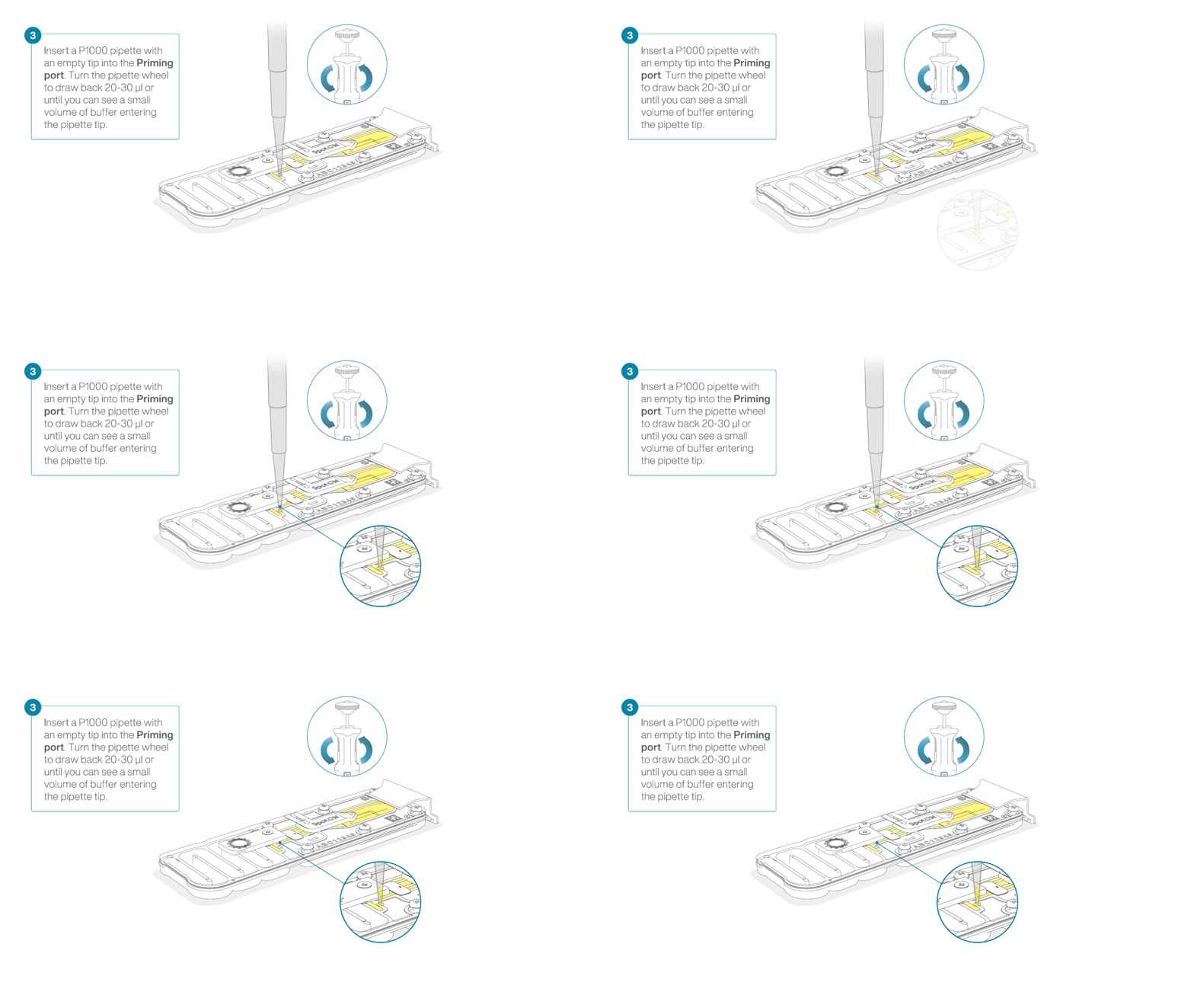
Load 800 µl of the priming mix into the flow cell via the priming port, avoiding the introduction of air bubbles. Wait for five minutes. During this time, prepare the library for loading by following the steps below.

Thoroughly mix the contents of the Library Beads (LIB) by pipetting.
The Library Beads (LIB) tube contains a suspension of beads. These beads settle very quickly. It is vital that they are mixed immediately before use.
We recommend using the Library Beads (LIB) for most sequencing experiments. However, the Library Solution (LIS) is available for more viscous libraries.
In a new 1.5 ml Eppendorf DNA LoBind tube, prepare the library for loading as follows:
| Reagent | Volume per flow cell |
|---|---|
| Sequencing Buffer (SB) | 37.5 µl |
| Library Beads (LIB) mixed immediately before use | 25.5 µl |
| DNA library | 12 µl |
| Total | 75 µl |
Complete the flow cell priming:
- Gently lift the SpotON sample port cover to make the SpotON sample port accessible.
- Load 200 µl of the priming mix into the flow cell priming port (not the SpotON sample port), avoiding the introduction of air bubbles.

Mix the prepared library gently by pipetting up and down just prior to loading.
Add 75 μl of the prepared library to the flow cell via the SpotON sample port in a dropwise fashion. Ensure each drop flows into the port before adding the next.

Gently replace the SpotON sample port cover, making sure the bung enters the SpotON port and close the priming port.

For optimal sequencing output, install the light shield on your flow cell as soon as the library has been loaded.
We recommend leaving the light shield on the flow cell when library is loaded, including during any washing and reloading steps. The shield can be removed when the library has been removed from the flow cell.
Place the light shield onto the flow cell, as follows:
Carefully place the leading edge of the light shield against the clip. Note: Do not force the light shield underneath the clip.
Gently lower the light shield onto the flow cell. The light shield should sit around the SpotON cover, covering the entire top section of the flow cell.

The MinION Flow Cell Light Shield is not secured to the flow cell and careful handling is required after installation.
Close the device lid and set up a sequencing run on MinKNOW.
When a flow cell is inserted into the MinION Mk1D, the device lid will sit on top of the flow cell, leaving a small gap around the sides. This is normal and has no impact on the performance of the device.
Please refer to this FAQ regarding the device lid.

7. Data acquisition and basecalling
We do not recommend sequencing and performing data analysis simultaneously on your device.
To ensure the compute on your device can keep up with the requirements for sequencing and/or analysis, we strongly recommend against running both processes at the same time.
Ensure your sequencing run has completed before setting off data analysis. Data analysis will be performed post-sequencing.
Equally, we do not recommend starting a sequencing run if you are currently performing data analysis on your device.
How to start sequencing
The MinKNOW software controls the sequencing device and carries out data acquisition and real-time basecalling. Please ensure MinKNOW is installed on your computer or device. Further instructions for setting up a sequencing run can be found in the MinKNOW protocol.
We recommend setting up your sequencing run using the basecalling and barcoding recommendations outlined below. All other parameters can be left to their default settings.
MinKNOW settings for rapid metagenomic sequencing
For the fastest turnaround time and easiest analysis, basecalling should be performed live during the sequencing run using the High Accuracy (HAC) basecaller.
Below are the recommendations for MinKNOW settings:
Positions
Flow cell position: [user defined]
Experiment name: [user defined]
Flow cell type: FLO-MIN114
Sample ID: [user defined]
Kit
Kit selection: Rapid PCR Barcoding Kit (SQK-RPB114-24)
Run configuration
Sequencing and analysis
Basecalling: On [default]
Modified bases: Off
Model: High-accuracy basecalling (HAC) [default]
Barcoding: On [default]
Trim barcodes: Off [default]
Barcode both ends: Off [default]
Custom barcodes selection: Off [default]
Alignment: Off [default]
Adaptive sampling: Off [default]
Advanced options
Active channel selection: On [default]
Time between pore scans: 1.5 [default]
Reserve pores: On [default]
Data targets
Run limit: [user defined]*
*Sequencing time will depend on data requirements. For rapid information, data can be analysed after as little as 30 minutes of sequencing. Or to maximise data generation, you can sequence for up to 72 hours.
Output
Output format
.POD5: On [default]
.FASTQ: On [default]
.BAM: On
Filtering: On [default]
Qscore: 9 [default]
Minimum read length: 200 bp [default]
8. Downstream analysis
We do not recommend sequencing and performing data analysis simultaneously on your device.
To ensure the compute on your device can keep up with the requirements for sequencing and/or analysis, we strongly recommend against running both processes at the same time.
Ensure your sequencing run has completed before setting off data analysis. Data analysis will be performed post-sequencing.
Equally, we do not recommend starting a sequencing run if you are currently performing data analysis on your device.
Post-basecalling analysis
We recommend performing downstream analysis using EPI2ME which facilitates bioinformatic analyses by allowing users to run Nextflow workflows in a desktop application. EPI2ME maintains a collection of bioinformatic workflows which are curated and actively maintained by experts in long-read sequence analysis.
Follow the instructions in the EPI2ME Installation guide to install the application on your device. For more information on how to use EPI2ME, refer to the EPI2ME Quick Start guide.
The basecalled data generated by the sequencing software can be easily analysed using the wf-metagenomics workflow, a bioinformatic pipeline written in Nextflow. This workflow provides identification and abundance estimation of taxa present in your sample.
Using the recommended database, human, fungal, bacterial, viral, archaea and protozoal sequences can be accurately identified using the workflow.
We recommend you always use the latest available version of the workflow. Further information about the usage and results provided by the workflow can be found in the wf-metagenomics EPI2ME documentation page.
Notes
You can also run this workflow through command line. However, we only recommend this option for experienced users. For more information and the latest version of the workflow, please visit the wf-metagenomics page on GitHub. If using the workflow through command line, the settings recommended for optimal workflow performance for this protocol are: --kraken2_confidence 0.01 --database_set PlusPF-8 --store_dir /path/to/database/download/directory/
/path/to/database/download/directory/ is a placeholder. The exact location used will depend on the user system.
Test data is available on Github to test the wf-metagenomics workflow.
The test data can be found in the following repository: wf-metagenomics test data.
Running wf-metagenomics with EPI2ME
Within the EPI2ME app, new workflows can be installed by selecting the appropriate workflow under the "Available Workflows" tab in the Workflows section.
Already installed workflows can be found under the "Installed" tab.
You can test your installation using a small test dataset that is provided with the workflow:
1. Launch the EPI2ME app.
2. Select View workflows.
3. Select the wf-metagenomics workflow.
4. Select Use demo data.
5. Launch the workflow.
6. Once the run has completed, results can be viewed under the "Report" tab. The workflow outputs an interactive HTML report.
Running the workflow
To run wf-metagenomics with the settings recommended for the dual arm metagenomics protocol:
1. Launch the EPI2ME app.
2. Select View workflows.
3. Select the wf-metagenomics workflow.
4. Select Run this workflow.
5. Choose local or cloud option (if available).
6. In "Input Options", specify the input data for the workflow (either FASTQ or BAM).
7. In "Sample Options", provide a sample sheet in comma-separated values (CSV) format.
We suggest you provide an alias for each barcode that describes both the sample and prep type (e.g. sample_A_viral, sample_A_bacterial).
8. In "Reference Options", choose PlusPF-8 database to ensure archaea, bacterial, viral, human, and fungal taxa can be identified by the workflow.
9. In "Kraken2 Options", set Confidence score threshold to 0.01.
10. Click Launch workflow.
11. Once the run has completed, results can be viewed under the "Report" tab. The workflow outputs an interactive HTML report.
Note: All options not specified should be left to their default values.
Optional: If you need to adjust the computational resource available to your workflow, you can change local CPU and memory allocation in the workflow setup.
9. Flow cell reuse and returns
材料
- Flow Cell Wash Kit (EXP-WSH004)
After your sequencing experiment is complete, if you would like to reuse the flow cell, please follow the Flow Cell Wash Kit protocol and store the washed flow cell at +2°C to +8°C.
The Flow Cell Wash Kit protocol is available on the Nanopore Community.
We recommend you to wash the flow cell as soon as possible after you stop the run. However, if this is not possible, leave the flow cell on the device and wash it the next day.
Alternatively, follow the returns procedure to send the flow cell back to Oxford Nanopore.
Instructions for returning flow cells can be found here.
If you encounter issues or have questions about your sequencing experiment, please refer to the Troubleshooting Guide that can be found in this protocol.
10. Issues during DNA/RNA extraction and library preparation
Below is a list of the most commonly encountered issues, with some suggested causes and solutions.
We also have an FAQ section available on the Nanopore Community Support section.
If you have tried our suggested solutions and the issue still persists, please contact Technical Support via email (support@nanoporetech.com) or via LiveChat in the Nanopore Community.
Low sample quality
| Observation | Possible cause | Comments and actions |
|---|---|---|
| Low DNA purity (Nanodrop reading for DNA OD 260/280 is <1.8 and OD 260/230 is <2.0–2.2) | The DNA extraction method does not provide the required purity | The effects of contaminants are shown in the Contaminants document. Please try an alternative extraction method that does not result in contaminant carryover. Consider performing an additional SPRI clean-up step. |
| Low RNA integrity (RNA integrity number <9.5 RIN, or the rRNA band is shown as a smear on the gel) | The RNA degraded during extraction | Try a different RNA extraction method. For more info on RIN, please see the RNA Integrity Number document. Further information can be found in the DNA/RNA Handling page. |
| RNA has a shorter than expected fragment length | The RNA degraded during extraction | Try a different RNA extraction method. For more info on RIN, please see the RNA Integrity Number document. Further information can be found in the DNA/RNA Handling page. We recommend working in an RNase-free environment, and to keep your lab equipment RNase-free when working with RNA. |
Low DNA recovery after AMPure bead clean-up
| Observation | Possible cause | Comments and actions |
|---|---|---|
| Low recovery | DNA loss due to a lower than intended AMPure beads-to-sample ratio | 1. AMPure beads settle quickly, so ensure they are well resuspended before adding them to the sample. 2. When the AMPure beads-to-sample ratio is lower than 0.4:1, DNA fragments of any size will be lost during the clean-up. |
| Low recovery | DNA fragments are shorter than expected | The lower the AMPure beads-to-sample ratio, the more stringent the selection against short fragments. Please always determine the input DNA length on an agarose gel (or other gel electrophoresis methods) and then calculate the appropriate amount of AMPure beads to use. 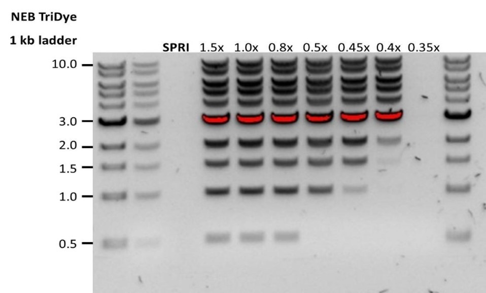 |
| Low recovery after end-prep | The wash step used ethanol <70% | DNA will be eluted from the beads when using ethanol <70%. Make sure to use the correct percentage. |
11. Issues during the sequencing run using a Rapid-based sequencing kit
Below is a list of the most commonly encountered issues, with some suggested causes and solutions.
We also have an FAQ section available on the Nanopore Community Support section.
If you have tried our suggested solutions and the issue still persists, please contact Technical Support via email (support@nanoporetech.com) or via LiveChat in the Nanopore Community.
Fewer pores at the start of sequencing than after Flow Cell Check
| Observation | Possible cause | Comments and actions |
|---|---|---|
| MinKNOW reported a lower number of pores at the start of sequencing than the number reported by the Flow Cell Check | An air bubble was introduced into the nanopore array | After the Flow Cell Check it is essential to remove any air bubbles near the priming port before priming the flow cell. If not removed, the air bubble can travel to the nanopore array and irreversibly damage the nanopores that have been exposed to air. The best practice to prevent this from happening is demonstrated in this video. |
| MinKNOW reported a lower number of pores at the start of sequencing than the number reported by the Flow Cell Check | The flow cell is not correctly inserted into the device | Stop the sequencing run, remove the flow cell from the sequencing device and insert it again, checking that the flow cell is firmly seated in the device and that it has reached the target temperature. If applicable, try a different position on the device (GridION/PromethION). |
| MinKNOW reported a lower number of pores at the start of sequencing than the number reported by the Flow Cell Check | Contaminations in the library damaged or blocked the pores | The pore count during the Flow Cell Check is performed using the QC DNA molecules present in the flow cell storage buffer. At the start of sequencing, the library itself is used to estimate the number of active pores. Because of this, variability of about 10% in the number of pores is expected. A significantly lower pore count reported at the start of sequencing can be due to contaminants in the library that have damaged the membranes or blocked the pores. Alternative DNA/RNA extraction or purification methods may be needed to improve the purity of the input material. The effects of contaminants are shown in the Contaminants Know-how piece. Please try an alternative extraction method that does not result in contaminant carryover. |
MinKNOW script failed
| Observation | Possible cause | Comments and actions |
|---|---|---|
| MinKNOW shows "Script failed" | Restart the computer and then restart MinKNOW. If the issue persists, please collect the MinKNOW log files and contact Technical Support. If you do not have another sequencing device available, we recommend storing the flow cell and the loaded library at 4°C and contact Technical Support for further storage guidance. |
Pore occupancy below 40%
| Observation | Possible cause | Comments and actions |
|---|---|---|
| Pore occupancy <40% | Not enough library was loaded on the flow cell | Ensure the correct concentration of good quality library is loaded on to a MinION/GridION flow cell. To check the concentration, please refer to the library preparation protocol. Please quantify the library before loading and calculate mols using tools like the Promega Biomath Calculator, choosing "dsDNA: µg to pmol" |
| Pore occupancy close to 0 | The Rapid Sequencing Kit V14/Rapid Barcoding Kit V14 was used, and sequencing adapters did not attach to the DNA | Make sure to closely follow the protocol and use the correct volumes and incubation temperatures. A Lambda control library can be prepared to test the integrity of reagents. |
| Pore occupancy close to 0 | No tether on the flow cell | Tethers are adding during flow cell priming (FCT tube). Make sure FCT was added to FCF before priming. |
Shorter than expected read length
| Observation | Possible cause | Comments and actions |
|---|---|---|
| Shorter than expected read length | Unwanted fragmentation of DNA sample | Read length reflects input DNA fragment length. Input DNA can be fragmented during extraction and library prep. 1. Please review the Extraction Methods in the Nanopore Community for best practice for extraction. 2. Visualise the input DNA fragment length distribution on an agarose gel before proceeding to the library prep. 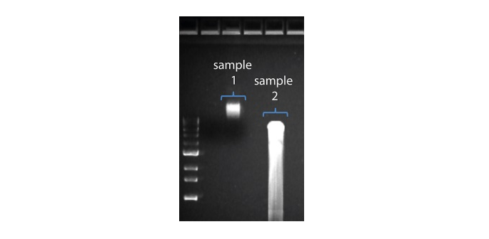 In the image above, Sample 1 is of high molecular weight, whereas Sample 2 has been fragmented. In the image above, Sample 1 is of high molecular weight, whereas Sample 2 has been fragmented.3. During library prep, avoid pipetting and vortexing when mixing reagents. Flicking or inverting the tube is sufficient. |
Large proportion of unavailable pores
| Observation | Possible cause | Comments and actions |
|---|---|---|
Large proportion of unavailable pores (shown as blue in the channels panel and pore activity plot) 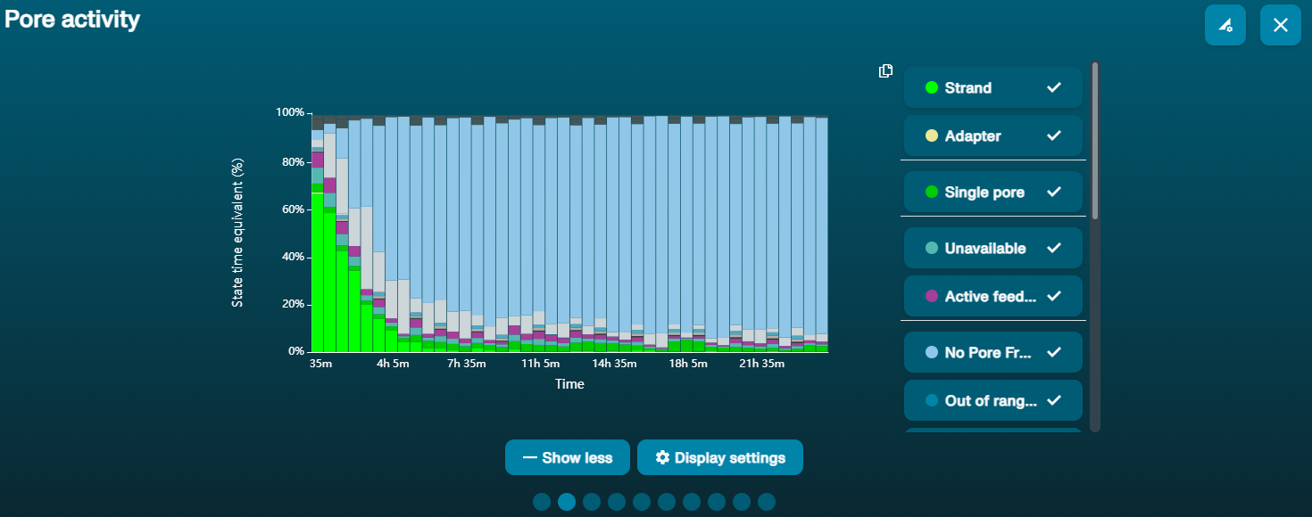 The pore activity plot above shows an increasing proportion of "unavailable" pores over time. The pore activity plot above shows an increasing proportion of "unavailable" pores over time. | Contaminants are present in the sample | Some contaminants can be cleared from the pores by the unblocking function built into MinKNOW. If this is successful, the pore status will change to "sequencing pore". If the portion of unavailable pores stays large or increases: 1. A nuclease flush using the Flow Cell Wash Kit (EXP-WSH004) can be performed, or 2. Run several cycles of PCR to try and dilute any contaminants that may be causing problems. |
Large proportion of inactive pores
| Observation | Possible cause | Comments and actions |
|---|---|---|
| Large proportion of inactive/unavailable pores (shown as light blue in the channels panel and pore activity plot. Pores or membranes are irreversibly damaged) | Air bubbles have been introduced into the flow cell | Air bubbles introduced through flow cell priming and library loading can irreversibly damage the pores. Watch the Priming and loading your flow cell video for best practice |
| Large proportion of inactive/unavailable pores | Certain compounds co-purified with DNA | Known compounds, include polysaccharides, typically associate with plant genomic DNA. 1. Please refer to the Plant leaf DNA extraction method. 2. Clean-up using the QIAGEN PowerClean Pro kit. 3. Perform a whole genome amplification with the original gDNA sample using the QIAGEN REPLI-g kit. |
| Large proportion of inactive/unavailable pores | Contaminants are present in the sample | The effects of contaminants are shown in the Contaminants Know-how piece. Please try an alternative extraction method that does not result in contaminant carryover. |
Temperature fluctuation
| Observation | Possible cause | Comments and actions |
|---|---|---|
| Temperature fluctuation | The flow cell has lost contact with the device | Check that there is a heat pad covering the metal plate on the back of the flow cell. Re-insert the flow cell and press it down to make sure the connector pins are firmly in contact with the device. If the problem persists, please contact Technical Services. |
Failed to reach target temperature
| Observation | Possible cause | Comments and actions |
|---|---|---|
| MinKNOW shows "Failed to reach target temperature" | The instrument was placed in a location that is colder than normal room temperature, or a location with poor ventilation (which leads to the flow cells overheating) | MinKNOW has a default timeframe for the flow cell to reach the target temperature. Once the timeframe is exceeded, an error message will appear and the sequencing experiment will continue. However, sequencing at an incorrect temperature may lead to a decrease in throughput and lower q-scores. Please adjust the location of the sequencing device to ensure that it is placed at room temperature with good ventilation, then re-start the process in MinKNOW. Please refer to this link for more information on MinION temperature control. |
