Multiplex methylation whole genome sequencing (MMWG_9241_v114_revA_11Nov2026)


PromethION: Protocol
Multiplex methylation whole genome sequencing V MMWG_9241_v114_revA_11Nov2026
This protocol provides details for library preparation, sequencing, and analysis overview.
The protocol:
- Uses genomic DNA extracted from tumour tissue sample
- Requires no PCR
- Is compatible with R10.4.1 flow cells
- Is for multiplexing four samples per PromethION Flow Cell
For Research Use Only
FOR RESEARCH USE ONLY.
Contents
Introduction to the protocol
- 1. Overview of the protocol
- 2. Guidance on recommended DNA extraction kits
- 3. Equipment and consumables
Sample preparation
Library preparation
- 6. DNA repair and end-prep
- 7. Native barcode ligation
- 8. Adapter ligation and clean-up
- 9. Priming and loading the PromethION Flow Cell
Sequencing and data analysis
Troubleshooting
概要
This protocol provides details for library preparation, sequencing, and analysis overview.
The protocol:
- Uses genomic DNA extracted from tumour tissue sample
- Requires no PCR
- Is compatible with R10.4.1 flow cells
- Is for multiplexing four samples per PromethION Flow Cell
For Research Use Only
1. Overview of the protocol
Introduction to the Multiplex methylation whole genome sequencing protocol
This protocol describes a nanopore sequencing method for low-pass whole genome sequencing to enable methylation-based classification of four tumour samples per PromethION Flow Cell. It is designed to generate sufficient genome-wide coverage for accurate methylation classification and arm-level copy number variation (CNV) while maximising sample throughput.
All Oxford Nanopore Technologies supplied reagents required for this protocol are included in the Multiplex Methylation Whole Genome Sequencing bundle available in 24-sample format. This bundle includes the Native Barcoding Kit and PromethION Flow Cells, and is the standard configuration for implementing this workflow.
Genomic DNA is extracted from fresh or frozen tumour tissue (or provided as purified DNA), and sequencing libraries are prepared using the Native Barcoding Kit (SQK-NBD114.24), allowing multiplexing of four samples per run on a flow cell. Barcoding enables robust sample tracking and demultiplexing to ensure accurate assignment of sequencing reads to each tumour sample.
Whole genome sequencing is performed for 16 hours, generating low-pass whole genome coverage suitable for methylation profiling and CNV calling. Analysis can be performed using the Somatic analysis bioinformatics guide which provides an overview of methods using third-party tools.
Overview of steps in the workflow
Prepare for your experiment
You will need to:
- Extract DNA from your input sample (e.g. tumour tissue).
- Ensure you have your sequencing kit, the correct equipment, and third-party reagents.
- Download the software for acquiring and analysing your data.
- Ensure your analysis workstation or compute environment meets the hardware and software requirements of your chosen third-party analysis tools.
- Check your flow cell to ensure it has sufficient pores for a good sequencing run.
Sample preparation, library preparation, and sequencing
| Step in workflow | Process | Time | Stop option |
|---|---|---|---|
| Sample preparation | Extract gDNA from sample and quantify. Fragment the gDNA and quantify. Check the length, quantity and purity of your extracted material. The quality checks performed during the protocol are recommended to ensure experimental success. | Dependent on extraction kit used ~15 minutes for shearing | At this stage, the extracted gDNA or fragmented gDNA can be stored at –20°C for later use. |
| DNA repair and end-prep | Repair the gDNA, and prepare the DNA ends for barcode and adapter attachment. | 60 minutes | 4°C overnight |
| Native barcode and adapter ligation | Ligate the native barcodes and sequencing adapters to the DNA ends. | 90 minutes | 4°C overnight |
| Priming and loading the flow cell | Prime the flow cell, and load your DNA library into the flow cell. | 10 minutes | |
| Sequencing | Sequence your library using your sequencing device. | 16 hours | |
| Data analysis | Refer to section 11, Downstream analysis and the Somatic analysis bioinformatics guide. |
Compatibility of this protocol
This protocol should only be used in combination with:
- Multiplex Methylation Whole Genome Sequencing bundle (MMWGS24)
- Native Barcoding Kit 24 V14 (SQK-NBD114.24)
- PromethION Flow Cell (FLO-PRO114M)
- PromethION 24: PromethION - device and IT specifications
- PromethION 2 Integrated: PromethION 2 Integrated - device and IT specifications
2. Guidance on recommended DNA extraction kits
DNA extraction and DNA input
Oxford Nanopore has tested and recommends the extraction kits for tumour tissue samples listed below, as well as the DNA input requirement.
| Tumour tissue (fresh or frozen) | |
|---|---|
| Kits | QIAamp Fast DNA Tissue Kit (Qiagen, 51404) Puregene Tissue Kit (Qiagen, 158063) MagAttract HMW DNA Kit (Qiagen, 67563) |
| DNA input per sample | 250 ng Up to 1 µg input is recommended where possible to ensure robust and consistent performance. |
Note: You will need to refer to the relevant kit handbook and protocol to determine which equipment and additional reagents are required for your extraction.
3. Equipment and consumables
材料
- 250 ng of extracted genomic DNA per sample
- Multiplex Methylation Whole Genome Sequencing bundle (containing the material listed below)
- Native Barcoding Kit 24 V14 (SQK-NBD114.24)
消耗品
- PromethION Flow Cell (FLO-PRO114M)
- g-TUBE (Covaris, 520079)
- Nuclease-free water (e.g. ThermoFisher, AM9937)
- Absolute ethanol (e.g. Sigma-Aldrich, 459836-100ML)
- NEB Blunt/TA Ligase Master Mix (NEB, M0367)
- NEBNext® Quick Ligation Reaction Buffer (NEB, B6058S)
- NEBNext® Companion Module v2 for Oxford Nanopore Technologies® Ligation Sequencing (NEB, E7672S or E7672L)
- Qubit™ dsDNA HS Assay Kit (Invitrogen, Q32851)
- Qubit™ Assay Tubes (Invitrogen, Q32856)
- 1.5 ml Eppendorf DNA LoBind tubes
- 0.2 ml 薄壁のPCRチューブ
- Pipette tips (nuclease-free, filtered recommended)
- Genomic DNA 165 kb Analysis Kit (Agilent, FP-1002-0275) (optional)
装置
- PromethION device
- Rotator mixer (e.g. Hula mixer)
- Magnetic separation rack, suitable for 1.5 ml Eppendorf tubes
- Eppendorf® 5424 centrifuge (or equivalent capable of ~3380 RCF)
- Microcentrifuge
- ボルテックスミキサー
- Thermal cycler with heated lid
- Heat block (suitable for 1.5 ml tubes)
- Qubit™ fluorometer (or equivalent for QC check)
- アイスバケツ(氷入り)
- タイマー
- P1000 pipette
- P200 pipette
- P100 pipette
- P20 pipette
- P10 pipette
- P2 pipette
オプション装置
- Femto Pulse System (Agilent Technologies) or equivalent
Oxford Nanopore Technologies Multiplex Methylation Whole Genome Sequencing bundle
All Oxford Nanopore Technologies reagents required for this protocol are included in the Multiplex Methylation Whole Genome Sequencing bundle, available in the Nanopore Store in 24-sample format.
This bundle includes the Native Barcoding Kit and PromethION Flow Cells, and is the standard configuration for implementing this workflow.
For more information on specific kit components found within this bundle, please refer to the relevant product found in the Nanopore Store page:
The above list of materials, consumables, and equipment is for the shearing of the DNA samples, as well as library preparation.
For genomic DNA extraction, we recommend using one of the extraction kits listed in section 2.
You will need to refer to the relevant kit handbook and protocol to determine which equipment and additional reagents are required for the extraction.
For this protocol, the following input is required for library preparation:
- 250 ng of sheared gDNA per sample
Input DNA
How to QC your input DNA
It is important that the input DNA meets the quantity and quality requirements. Using too little or too much DNA, or DNA of poor quality (e.g. highly fragmented or containing RNA or chemical contaminants) can affect your library preparation.
For instructions on how to perform quality control of your DNA sample, please read the Input DNA/RNA QC protocol.
Chemical contaminants
Depending on how the DNA is extracted from the raw sample, certain chemical contaminants may remain in the purified DNA, which can affect library preparation efficiency and sequencing quality. Read more about contaminants on the Contaminants page of the Community.
Third-party reagents
We have validated and recommend the use of all the third-party reagents used in this protocol. Alternatives have not been tested by Oxford Nanopore.
For all third-party reagents, we recommend following the manufacturer's instructions to prepare the reagents for use, unless specified in this protocol.
Check your flow cell
We highly recommend that you check the number of pores in your flow cell prior to starting a sequencing experiment, within the warranty period. Flow cells should be removed from refrigeration and their packaging and allowed to come to room temperature for 30 minutes before they are used.
To perform the flow cell check, please follow the instructions in the Flow Cell Check document. As part of your flow cell check, you will see the number of pores in your new flow cell. The warranty on your flow cell covers a minimum pore count of 5,000 pores for a PromethION Flow Cell. You can find more information about the warranty period, stability and storage conditions of flow cells on the Oxford Nanopore Store. If you have any concerns or questions, please contact Oxford Nanopore.
Oxford Nanopore will replace any unused PromethION Flow Cell with fewer than 5,000 pores, when the result is reported within two days of performing the flow cell check, and when the storage recommendations have been followed.
The Native Adapter (NA) used in this protocol is not interchangeable with other sequencing adapters.
4. Genomic DNA extraction
Extract genomic DNA from your sample using a recommended kit listed in section 2, following the manufacturer's instructions.
Following extraction, quantify 1 μl of extracted gDNA using the Qubit dsDNA HS Assay Kit following the manufacturer's instructions.
Optional: If interested in fragment size distribution analysis, reserve an additional 1 μl aliquot of the unsheared DNA for analysis on the Femto Pulse System. This should be run after sequencing to avoid increasing library preparation times.
Optional DNA QC
Tumour tissue
Following gDNA extraction, samples may be assessed on the Femto Pulse System to visualise the unsheared gDNA profiles. The comparison with representative traces from samples that did or did not subsequently yield acceptable unsheared fragment distributions can be used to guide QC (Figure 1).
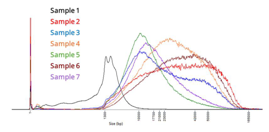
Figure 1: Unsheared genomic DNA fragment length profiles from seven commercial tumour tissue samples extracted using the QIAamp Fast DNA Tissue Kit. Samples were assessed using the Agilent Femto Pulse System with the Genomic DNA 165 kb Kit.
Sample 1 displayed reduced fragment lengths consistent with higher levels of necrosis, while Samples 2–7 demonstrated acceptable fragment size distributions.
Take forward your extracted gDNA into the next step, DNA shearing.
Alternatively, your sample(s) can be stored at 4°C overnight or at –20°C for later use.
5. DNA shearing using g-TUBE
材料
- 250 ng extracted gDNA per each of the 4 samples
消耗品
- Four g-TUBEs (Covaris, 520079) (one per sample)
- Nuclease-free water (e.g. ThermoFisher, AM9937)
- Qubit™ dsDNA HS Assay Kit (Invitrogen, Q32851)
- Qubit™ Assay Tubes (Invitrogen, Q32856)
- 1.5 ml Eppendorf DNA LoBind tubes
- Pipette tips (nuclease-free, filtered recommended)
- Genomic DNA 165 kb Analysis Kit (Agilent, FP-1002-0275) (optional)
装置
- Eppendorf® 5424 centrifuge (or equivalent capable of ~3380 RCF)
- Microcentrifuge
- Qubit™ fluorometer (or equivalent for QC check)
- アイスバケツ(氷入り)
- P1000 pipette
- P200 pipette
- P100 pipette
- P20 pipette
- P10 pipette
- P2 pipette
オプション装置
- Femto Pulse System (Agilent Technologies) or equivalent
To fragment gDNA, dilute 250 ng of sample DNA in nuclease-free water to a total volume of 52 μl. Dilute the four samples in separate 1.5 ml Eppendorf tubes.
Load the four diluted samples into four different g-TUBEs according to the manufacturer’s instructions. Balance the tubes in the centrifuge appropriately.
Centrifuge the g-TUBEs at ~3,381 RCF (6,000 RPM) for 30 seconds. If using an odd number of samples, balance the centrifuge with an additional tube.
Visually inspect the g-TUBEs to confirm that the samples have moved completely from the upper to the lower chambers. If they have, invert the g-TUBEs and centrifuge at ~3,381 RCF (6,000 RPM) for 30 seconds.
If the liquid has not passed into the lower chamber completely, do not invert the tube and centrifuge for an additional 30 seconds at ~3,381 RCF (6,000 RPM) to ensure that all material has passed through the gTUBE constriction. Once completed, invert the g-TUBE and continue.
Repeat steps 3 and 4 to perform a second pass of shearing.
With the sample volume in the lower chamber, unscrew the g-TUBE body, leaving the screw-cap containing the sample volume.
Retrieve each sample from the g-TUBE screw-cap and transfer it into separate, clean 1.5 ml Eppendorf tube.
Quantify 1 μl of sheared DNA using the Qubit dsDNA HS Assay Kit following the manufacturer's instructions.
The minimum concentration should be ~5 ng/μl depending on the input used.
Optional: If interested in fragment size distribution analysis, reserve an additional 1 μl aliquot of the sheared DNA for analysis on the Femto Pulse System. This should be run after sequencing to avoid increasing library preparation times.
Optional DNA QC
Tumour tissue
Following shearing, samples may be assessed on the Femto Pulse System to visualise the sheared gDNA profiles. The comparison with representative traces from samples that did or did not subsequently yield acceptable sheared fragment distributions can be used to guide QC (Figure 2).
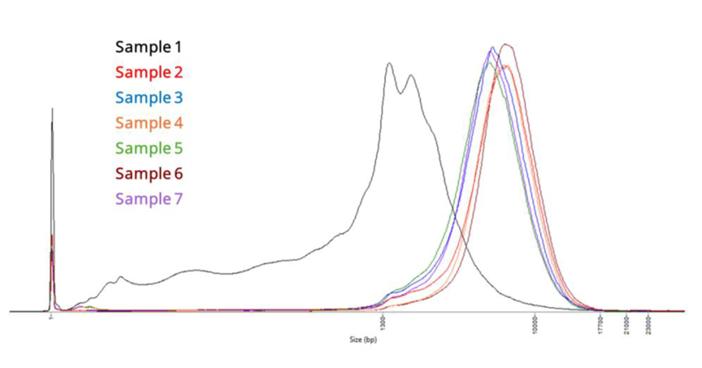
Figure 2: Sheared genomic DNA fragment length profiles from seven commercial tumour tissue samples extracted using the QIAamp Fast DNA Tissue Kit followed by g-TUBE shearing. Samples were assessed using the Agilent Femto Pulse System with the Genomic DNA 165 kb Kit.
Sample 1 exhibited reduced fragment lengths after shearing, consistent with increased necrosis, whereas Samples 2–7 showed acceptable fragment size distributions. Post-shearing showed a modal fragment size of approximately 10 kb.
Take forward a minimum of 250 ng of sheared DNA for each of the four samples into the library preparation section.
6. DNA repair and end-prep
材料
- 250 ng sheared DNA per sample (from previous step)
- AMPure XP Beads (AXP from the Native Barcoding Kit 24 V14)
消耗品
- Nuclease-free water (e.g. ThermoFisher, AM9937)
- Absolute ethanol (e.g. Sigma-Aldrich, 459836-100ML)
- NEBNext® Companion Module v2 for Oxford Nanopore Technologies® Ligation Sequencing (NEB, E7672S or E7672L)
- Qubit™ dsDNA HS Assay Kit (Invitrogen, Q32851)
- Qubit™ Assay Tubes (Invitrogen, Q32856)
- 1.5 ml Eppendorf DNA LoBind tubes
- 0.2 ml 薄壁のPCRチューブ
- Pipette tips (nuclease-free, filtered recommended)
装置
- Thermal cycler with heated lid
- Microcentrifuge
- ボルテックスミキサー
- Rotator mixer (e.g. Hula mixer)
- Magnetic separation rack, suitable for 1.5 ml Eppendorf tubes
- Heat block (suitable for 1.5 ml tubes) set to 37°C
- Qubit™ fluorometer (or equivalent for QC check)
- タイマー
- アイスバケツ(氷入り)
- P1000 pipette
- P200 pipette
- P100 pipette
- P20 pipette
- P10 pipette
- P2 pipette
Prepare the required reagents from the NEBNext Comapnion Module v2 in accordance with the manufacturer’s instructions.
To a 0.2 ml PCR tube, add the following reagents for each sample:
| Reagent | Volume |
|---|---|
| 250 ng sheared input DNA (from previous step) | 48 µl |
| NEBNext® FFPE DNA Repair Buffer v2 | 7 µl |
| NEBNext® FFPE DNA Repair Mix | 2 µl |
| NEBNext® Ultra™ II End Prep Enzyme Mix | 3 µl |
| Total | 60 µl |
Optional:
The FFPE DNA Repair Buffer v2, FFPE DNA Repair Mix and Ultra™ II End Prep Enzyme Mix can be prepared as a mastermix for four samples (including excess) and 12 µl aliquoted into 0.2 ml PCR tubes.
Then add 48 μl of sheared DNA to each tube of mastermix.
Mix the components thoroughly by pipetting and spin down briefly.
Incubate the reactions in a thermal cycler at 20°C for 5 minutes, then 65°C for 5 minutes.
Cool the reactions briefly on ice.
Note: This is to ensure that the reactions are at room temperature before the addition of the AMPure XP beads.
Spin down and transfer each sample to a clean 1.5 ml Eppendorf tube and add 60 μl (1x ratio) of resuspended AMPure XP beads.
Pipette mix and incubate the samples on a rotator mixer (e.g. Hula mixer) at room temperature for 5 minutes.
Freshly prepare sufficient 80% ethanol in nuclease-free water for the number of samples being prepped.
Spin down each sample and pellet on a magnetic rack for at least 1–3 minutes, until the eluate is clear and colourless. Keep the tubes on the magnetic rack and pipette off the supernatants.
Keep the tubes on the magnetic rack and wash the beads with 200 µl of freshly prepared 80% ethanol, without disturbing the pellets. Remove the supernatants using a pipette and discard.
Repeat the previous step.
Spin down and place the samples back on the magnetic rack. Pipette off any residual supernatants. Allow the pellets to dry for ~30 seconds, but do not dry the pellets to the point of cracking.
Remove the samples from the magnetic rack and resuspend each pellet in 11 μl of nuclease-free water.
Incubate the samples on a heat block set at 37°C for 2 minutes with intermittent flicking of the tubes to aid DNA elution from the beads.
Spin down the samples and pellet on the magnetic rack for at least 1–3 minutes, until the eluates are clear and colourless.
Collect and retain each eluate in separate, clean 1.5 ml Eppendorf tubes.
Quantify the eluates using the Qubit dsDNA HS Assay Kit following the manufacturer's instructions.
The expected minimum concentration for the eluate is ~14 ng/μl.
Take forward your end-prepped DNA samples into the native barcode ligation step.
7. Native barcode ligation
材料
- Reagents from the Native Barcoding Kit 24 V14 (SQK-NBD114.24):
- Native Barcodes (NB01-24)
- EDTA (EDTA)
- AMPure XP Beads (AXP)
- Short Fragment Buffer (SFB)
消耗品
- NEB Blunt/TA Ligase Master Mix (NEB, M0367)
- Nuclease-free water (e.g. ThermoFisher, AM9937)
- Qubit™ dsDNA HS Assay Kit (Invitrogen, Q32851)
- Qubit™ Assay Tubes (Invitrogen, Q32856)
- 1.5 ml Eppendorf DNA LoBind tubes
- Pipette tips (nuclease-free, filtered recommended)
装置
- Microcentrifuge
- ボルテックスミキサー
- Rotator mixer (e.g. Hula mixer)
- Magnetic separation rack, suitable for 1.5 ml Eppendorf tubes
- Heat block (suitable for 1.5 ml tubes) set to 37°C
- Qubit™ fluorometer (or equivalent for QC check)
- タイマー
- アイスバケツ(氷入り)
- P1000 pipette
- P200 pipette
- P100 pipette
- P20 pipette
- P10 pipette
- P2 pipette
Prepare the required reagents from NEB in accordance with the manufacturer’s instructions.
Thaw the Native Barcodes (NB01-24) at room temperature . Individually mix the barcodes by pipetting, spin down, and place them on ice.
The wells of the barcoding plate are intended for single use only. Please ensure your barcode well is sealed before use. Do not reuse the barcode wells.
In a 1.5 ml Eppendorf tube, add the reagents in the following order for each of the four barcoding reactions:
Note: Only use one unique barcode for each of the four samples.
| Reagent | Volume |
|---|---|
| End-prepped DNA | 7.5 µl |
| Native Barcode (NB01-24) | 2.5 µl |
| Blunt/TA Ligase Master Mix | 10 µl |
| Total | 20 µl |
Mix well by pipetting and spin down the reactions. Incubate at room temperature for 20 minutes.
Add 4 µl of EDTA to each barcoding reaction and mix thoroughly by pipetting and spin down briefly.
Pool the four barcoded samples into a 1.5 ml Eppendorf DNA LoBind tube.
Note: For four samples, the total volume should be ~96 µl.
To the above reaction, add 38.4 µl (0.4x) of resuspended AMPure XP beads and incubate on a rotator mixer at room temperature for 10 minutes.
Spin down and place the sample tube on the magnetic rack. Allow the beads to pellet for at least 3 minutes. Keep the tube on the magnetic rack and pipette off the supernatant.
Wash the beads by adding 500 μl of S Fragment Buffer (SFB). Fully resuspend the pellet by flicking the tube. Pellet the beads on the magnetic rack for at least 3–5 minutes, until the supernatant is clear. Remove the supernatant using a pipette and discard.
Repeat the previous step.
Spin down and place the sample tube back on the magnetic rack. Pipette off any residual supernatant. Allow the pellet to dry for ~30 seconds, but do not dry the pellet to the point of cracking.
Remove the sample tube from the magnetic rack and resuspend the pellet in 32 μl of nuclease-free water.
Spin down the sample tube and incubate in a heat block set at 37°C for 10 minutes. Every 2 minutes, agitate the sample tube by gently flicking for 10 seconds to aid DNA elution.
Pellet the beads on the magnetic rack for at least 1–3 minutes, until the eluate is clear and colourless.
Remove and retain 31 μl of eluate containing the DNA library into a clean 1.5 ml Eppendorf DNA LoBind tube.
Quantify 1µl of eluate using the Qubit dsDNA HS Assay Kit following the manufacturer's instructions.
The expected minimum concentration is ~10 ng/µl.
Take forward the barcoded DNA library into the adapter ligation step.
8. Adapter ligation and clean-up
材料
- Reagents from the Native Barcoding Kit 24 V14 (SQK-NBD114.24):
- AMPure XP Beads (AXP)
- Native Adapter (NA)
- Long Fragment Buffer (LFB)
- Elution Buffer (EB)
消耗品
- NEBNext® Quick Ligation Reaction Buffer (NEB, B6058S)
- NEBNext® Companion Module v2 for Oxford Nanopore Technologies® Ligation Sequencing (NEB, E7672S or E7672L)
- Qubit™ dsDNA HS Assay Kit (Invitrogen, Q32851)
- Qubit™ Assay Tubes (Invitrogen, Q32856)
- 1.5 ml Eppendorf DNA LoBind tubes
- Pipette tips (nuclease-free, filtered recommended)
装置
- Microcentrifuge
- ボルテックスミキサー
- Rotator mixer (e.g. Hula mixer)
- Magnetic separation rack, suitable for 1.5 ml Eppendorf tubes
- Heat block (suitable for 1.5 ml tubes) set to 37°C
- Qubit™ fluorometer (or equivalent for QC check)
- タイマー
- アイスバケツ(氷入り)
- P1000 pipette
- P200 pipette
- P100 pipette
- P20 pipette
- P10 pipette
- P2 pipette
The Native Adapter (NA) used in this protocol is not interchangeable with other sequencing adapters.
Check your flow cell
We recommend performing a flow cell check before starting adapter ligation to ensure you have a flow cell with sufficient pores for a good sequencing run.
Flow cells should be removed from refrigeration and their packaging and allowed to come to room temperature for 30 minutes prior to use.
Condensation can form on the flow cells in humid environments. Inspect the gold connector pins on the top and underside of the flow cell for condensation and wipe off with a lint-free wipe if any is observed. Ensure the heat pad (dark grey) is present on the underside of the flow cell.
Refer to the flow cell check document for more information.
Prepare the NEBNext Quick Ligation Reaction Buffer and Salt-T4 DNA Ligase according to the manufacturer's instructions.
Add the reagents in the following order for the adapter ligation reaction into a 1.5 ml Eppendorf tube:
| Reagent | Volume |
|---|---|
| End-prepped and barcode ligated DNA | 30 µl |
| Native Adapter (NA) | 5 µl |
| NEBNext® Quick Ligation Reaction Buffer (5X) | 10 µl |
| Salt-T4® DNA Ligase | 5 µl |
| Total | 50 µl |
Thoroughly mix the reaction by gently pipetting and briefly spinning down.
Incubate the reaction for 20 minutes at room temperature.
Resuspend the AMPure XP beads by vortexing.
Add 20 µl (0.4x) of resuspended AMPure XP beads to the reaction and mix by pipetting.
Incubate on a rotator mixer for 10 minutes at room temperature.
Spin down the sample and pellet on the magnetic rack for at least 3 minutes until the beads have pelleted. Keep the tube on the magnetic rack and pipette off the supernatant.
Wash the beads by adding 125 μl of L Fragment Buffer (LFB). Flick the sample tube to resuspend the beads, then spin down. Return the tube to the magnetic rack and allow the beads to pellet for at least 3–5 minutes. Remove the supernatant using a pipette and discard.
Repeat the previous step.
Spin down and place the sample tube back on the magnetic rack. Pipette off any residual supernatant. Allow the pellet to dry for ~30 seconds, but do not dry the pellet to the point of cracking.
Remove the sample tube from the magnetic rack and resuspend the pellet in 34 µl of Elution Buffer (EB).
Spin down the sample and incubate in a heat block set at 37°C for 10 minutes. Every 2 minutes, agitate the sample by gently flicking the tube for 10 seconds to aid DNA elution.
Pellet the beads on the magnetic rack for at least 1 minute, until the eluate is clear and colourless.
Remove and retain 33 µl of eluate containing the DNA library into a clean 1.5 ml Eppendorf DNA LoBind tube.
Quantify 1µl of eluate using the Qubit dsDNA HS Assay Kit following the manufacturer's instructions.
The expected minimum concentration is ~3 ng/µl.
The prepared library is used for loading onto the flow cell. Store the library on ice or at 4°C until ready to load.
9. Priming and loading the PromethION Flow Cell
材料
- Reagents from the Native Barcoding Kit 24 V14 (SQK-NBD114.24):
- Flow Cell Flush (FCF)
- Flow Cell Tether (FCT)
- Library Beads (LIB)
- Sequencing Buffer (SB)
消耗品
- PromethION Flow Cell (FLO-PRO114M)
- 1.5 ml Eppendorf DNA LoBind tubes
- Pipette tips (nuclease-free, filtered recommended)
装置
- PromethION device
- PromethION Flow Cell Light Shield
- Microcentrifuge
- ボルテックスミキサー
- タイマー
- アイスバケツ(氷入り)
- P1000 pipette
- P200 pipette
- P100 pipette
- P20 pipette
This kit is only compatible with R10.4.1 flow cells (FLO-PRO114M).
We recommend removing your flow cells from refrigeration and their packaging, and allowing them to come to room temperature for 30 minutes before they are inserted into the device.
Condensation can form on the flow cell in humid environments. Inspect the gold connector pins on the top and underside of the flow cell for condensation and wipe off with a lint-free wipe if any is observed. Ensure the heat pad (dark grey) is present on the underside of the flow cell.
Thaw the Sequencing Buffer (SB), Library Beads (LIB), Flow Cell Tether (FCT) and Flow Cell Flush (FCF) at room temperature before mixing by vortexing. Then spin down and store on ice.
Prepare the flow cell priming mix in a suitable tube for the number of flow cells required. Once combined, mix well by briefly vortexing and spin down.
| Reagent | Volume per flow cell |
|---|---|
| Flow Cell Tether (FCT) | 30 µl |
| Flow Cell Flush (FCF) | 1,170 µl |
| Total volume | 1,200 µl |
Load the flow cell(s) into the device docking ports.
Make sure that the dark grey heat pad on the underside of the flow cell is not torn or deformed.
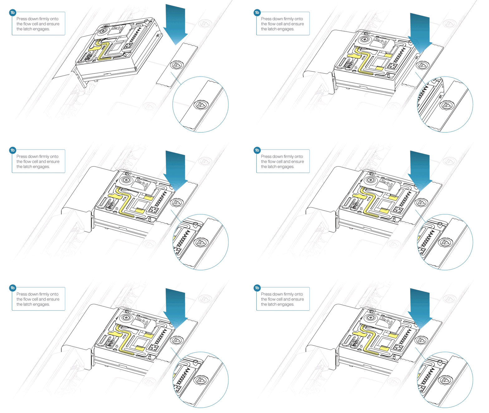
For the PromethION 24:
- Line up the flow cell with the connector horizontally and vertically before smoothly inserting into position.
- Press down firmly onto the flow cell and ensure the latch engages and clicks into place.
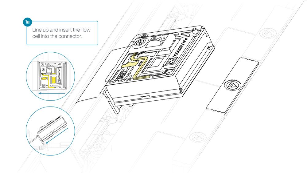
For the PromethION 2 Integrated:
Place the flow cell flat on the metal plate.
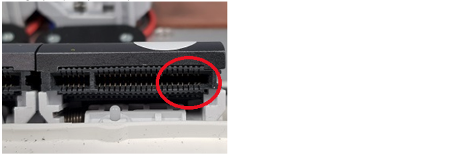
Slide the flow cell into the docking port until the gold pins cannot be seen.
Press the flow cell firmly into the loader until a click is heard.

Insertion of the flow cells at the wrong angle can cause damage to the pins on the PromethION and affect your sequencing results. If you find the pins on a PromethION position are damaged, please contact support@nanoporetech.com for assistance.
Complete a flow cell check to assess the number of pores available before loading the library.
This step can be omitted if the flow cell has been checked previously.
Refer to the flow cell check document for more information.
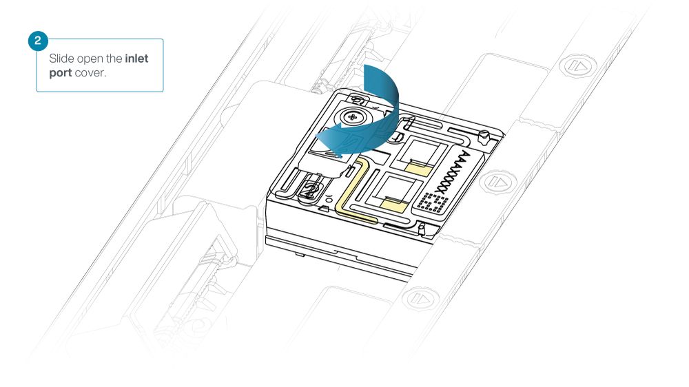
Slide the inlet port cover clockwise to open.
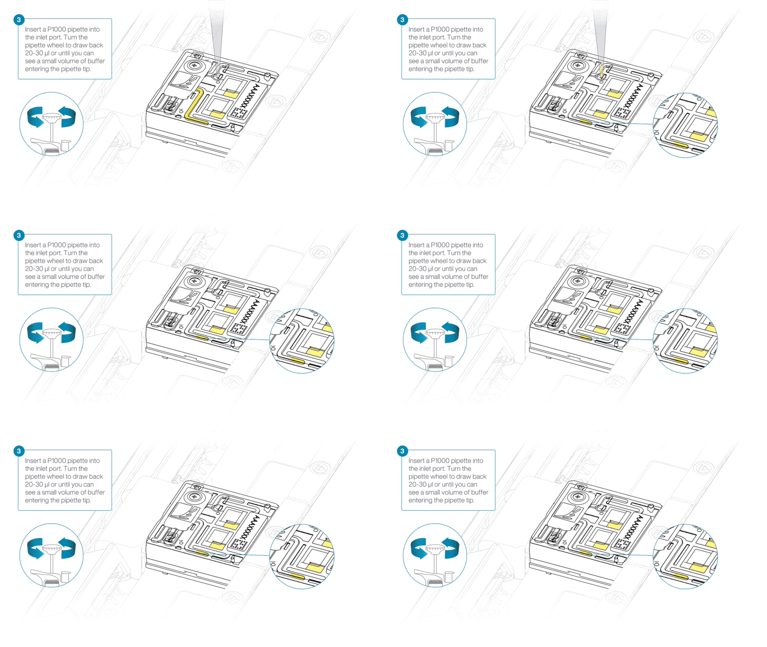
Take care when drawing back buffer from the flow cell. Do not remove more than 20–30 µl, and make sure that the array of pores are covered by buffer at all times. Introducing air bubbles into the array can irreversibly damage pores.
After opening the inlet port, draw back a small volume to remove any air bubbles:
- Set a P1000 pipette tip to 200 µl.
- Insert the tip into the inlet port.
- Turn the wheel until the dial shows 220–230 µl, or until you see a small volume of buffer entering the pipette tip.
Load 500 µl of the priming mix into the flow cell via the inlet port, avoiding the introduction of air bubbles. Wait five minutes. During this time, prepare the library for loading using the next steps in the protocol.
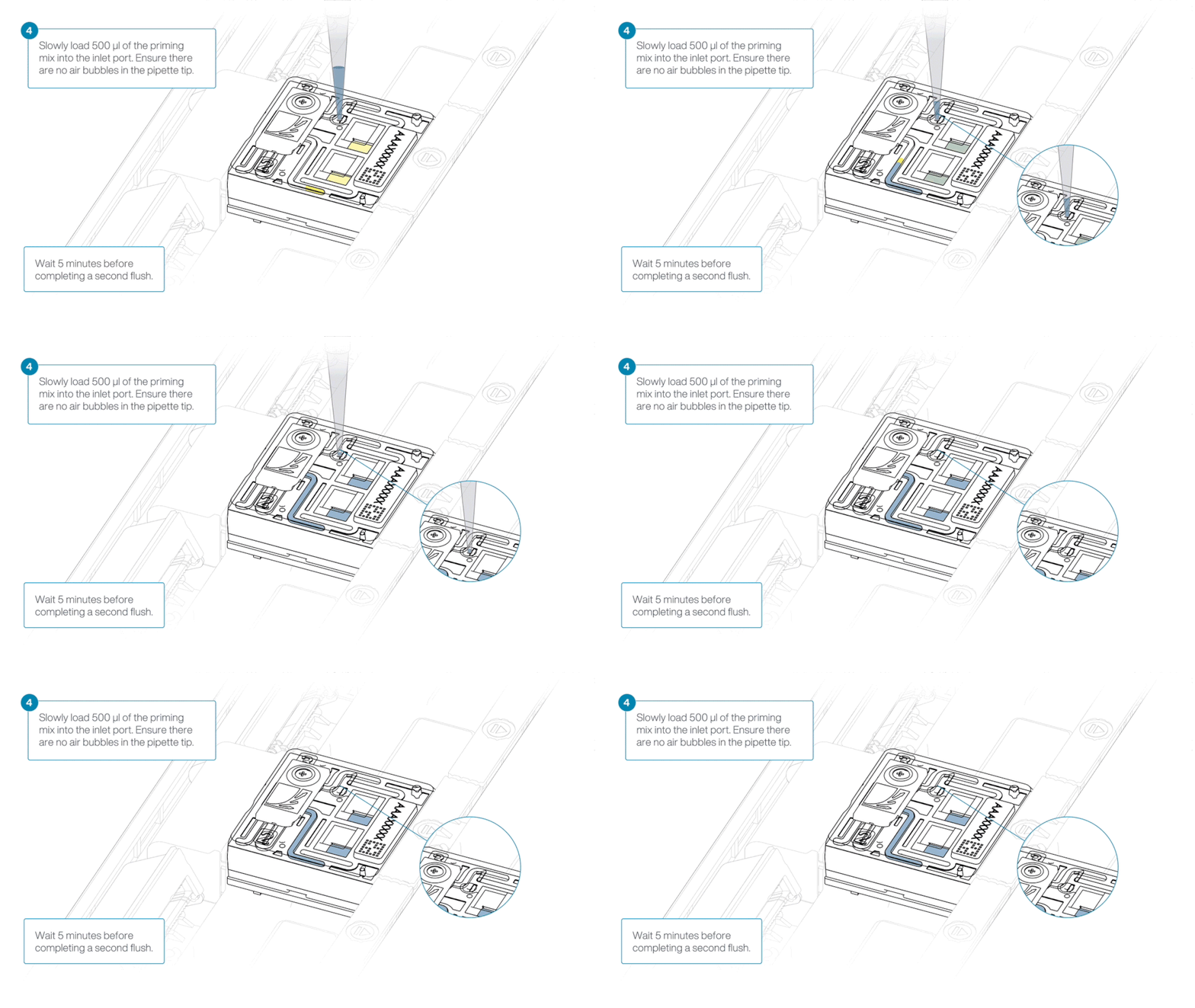
Thoroughly mix the contents of the Library Beads (LIB) by pipetting.
The Library Beads (LIB) tube contains a suspension of beads. These beads settle very quickly. It is vital that they are mixed immediately before use.
In a new 1.5 ml Eppendorf DNA LoBind tube, prepare the library for loading as follows:
| Reagent | Volume per flow cell |
|---|---|
| Sequencing Buffer (SB) | 100 µl |
| Library Beads (LIB) thoroughly mixed before use | 68 µl |
| DNA library | 32 µl |
| Total | 200 µl |
Complete the flow cell priming by slowly loading 500 µl of the priming mix into the inlet port.
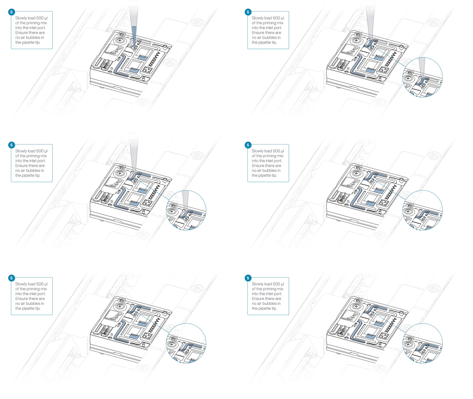
Mix the prepared library gently by pipetting up and down just prior to loading.
Load 200 µl of library into the inlet port using a P1000 pipette.
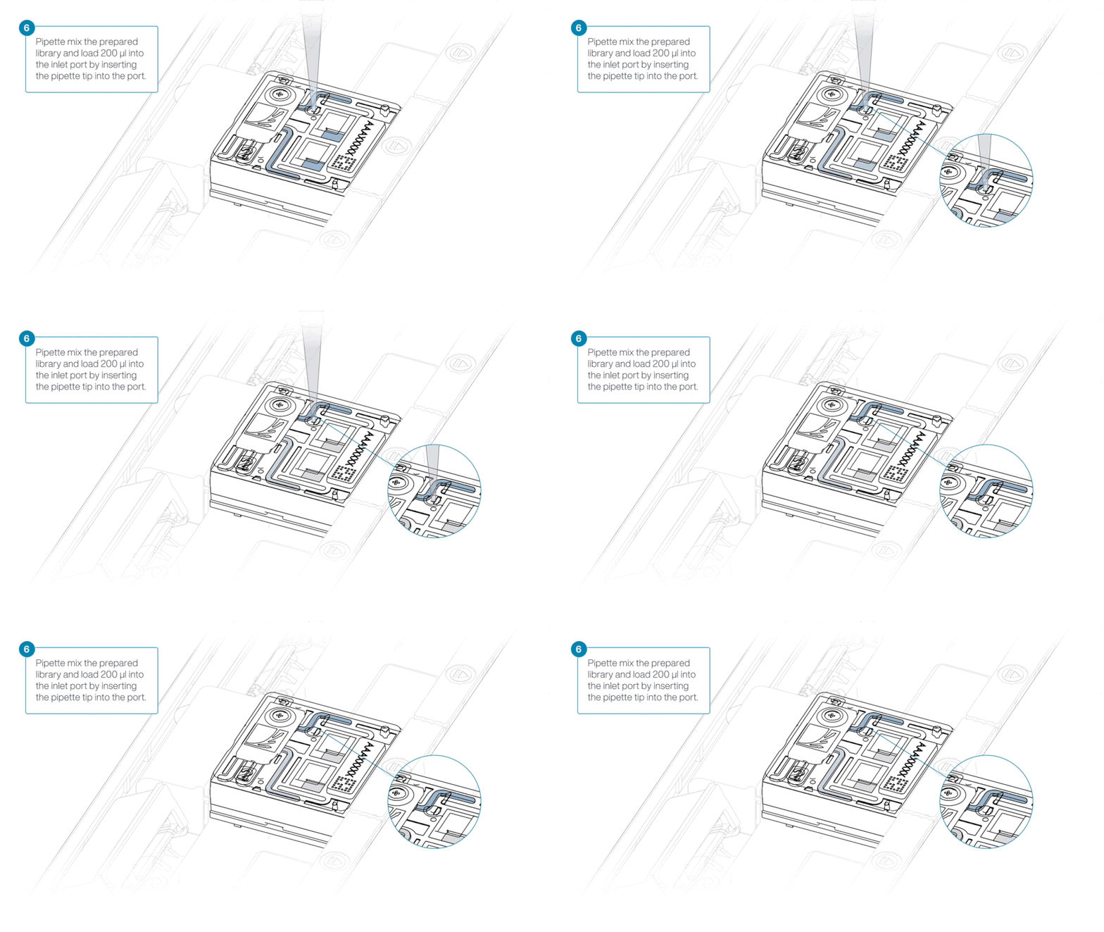
Close the valve to seal the inlet port.

For optimal sequencing output, install the light shield on your flow cell as soon as the library has been loaded.
We recommend leaving the light shield on the flow cell when library is loaded, including during any washing and reloading steps. The shield can be removed when the library has been removed from the flow cell.
If the light shield has been removed from the flow cell, install the light shield as follows:
- Align the inlet port cut out of the light shield with the inlet port cover on the flow cell. The leading edge of the light shield should sit above the flow cell ID.
- Firmly press the light shield around the inlet port cover. The inlet port clip will click into place underneath the inlet port cover.
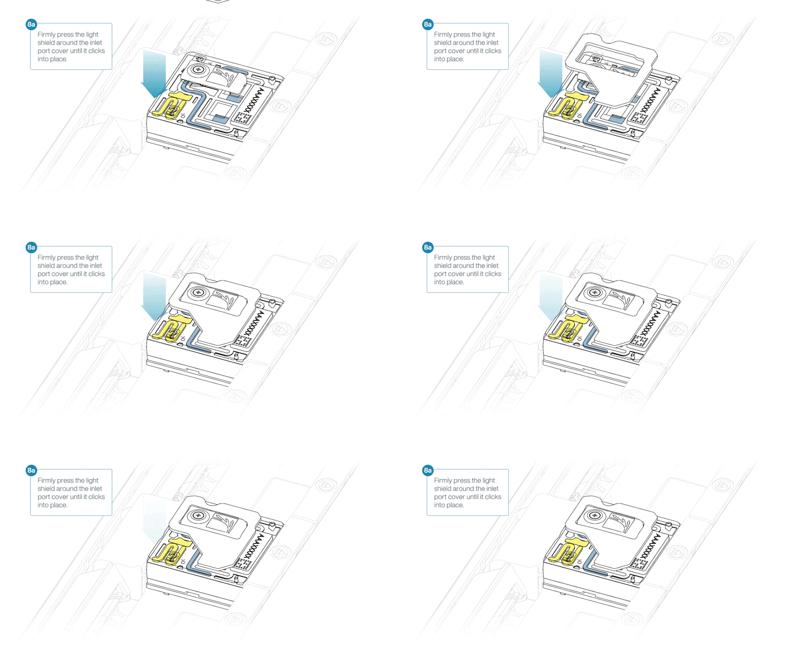
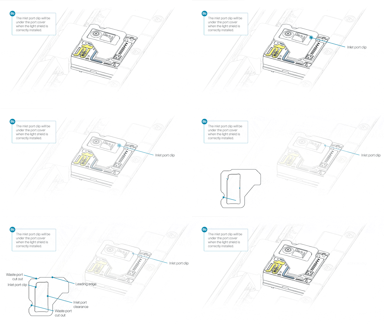
Close the PromethION lid when ready to start a sequencing run on MinKNOW.
Wait a minimum of 10 minutes after loading the library and before initiating any experiments. This will help to increase the sequencing output.
10. Data acquisition and basecalling
How to start sequencing
The sequencing device control, data acquisition and real-time basecalling are carried out by the MinKNOW software.
We recommend basecalling with the high accuracy (HAC) basecaller in real-time with BAM selected as output type using the PromethION 24 (P24) or PromethION 2 Integrated (P2i) device.
Refer to the links below containing the detailed instructions for setting up the device and sequencing run:
- PromethION 24: Starting a sequencing run on PromethION 24
- PromethION 2 Integrated: Starting a sequencing run on PromethION 2 Integrated
Below are the recommended sequencing parameters for MinKNOW.
Setting up your sequencing run in MinKNOW
Open the MinKNOW software UI via your desktop application.
In the MinKNOW UI, choose Start sequencing.
Follow the sequencing run set up on-screen using the settings defined below.
Positions
- Flow cell position: [user defined]
- Experiment name: [user defined]
- Flow cell type: FLO-PRO114M
- Sample ID: [user defined]
Kit selection
- Kit selection: Native Barcoding Sequencing Kit (SQK-NBD114)
Run configuration
- Sequencing and analysis
- Basecalling: On [default]
- Modified bases: On (5hmC and 5mC in CG contexts)
- Model: High accuracy basecalling (HAC)
- Barcoding: On [default]
Alignment: On
- Reference for alignment: GRCh38_GIABv3
Adaptive sampling: Off
Data targets
- Run limit: Stop when sequencing reaches 16 h
Output Settings
- Saved output file location: remote location for third-party methylation classification analysis or local directory [user defined]
Output format
- POD5: Off
Note: Enable POD5 output if downstream re-basecalling is anticipated - FASTQ: Off
- BAM: On [default]
- Based on: Time elapsed
- Frequency: Every 1 hour [default]
- POD5: Off
When performing methylation-based classification, consult the requirements of your chosen third-party classifier regarding recommended BAM file size.
The MinKNOW software can generate BAM files based on either a specified time interval or a predefined read count, providing flexibility to accommodate different analysis workflows. As methylation classifiers may have different input requirements, ensure that the BAM file generation settings are configured accordingly.
11. Downstream analysis
Methylation classification
Methylation classification using third-party tools can be performed after 2 hours of sequencing, or once approximately 0.75 Gb of pass data has been generated per sample.
Methylation calls are contained within the BAM files generated by MinKNOW, which are saved in the run output directory:
/data/{experiment_group}/{sample_id}/{start_time}_{device_id}_{flow_cell_id}_{short_run_id}/bam_pass/
Example:
/data/TMP_Verification/VQL/20260514_1430_P2S-01234-_PAA12345_e05460c6/bam_pass/
Copy number analysis
CNV calling can be performed after 16 hours of sequencing, when each sample is expected to generate sufficient data, with target outputs of >6 Gb (>2x) of PASS data per barcode.
The Somatic analysis bioinformatics guide provides an overview of methods for the analysis.
12. Flow cell reuse and returns
We do not recommend washing and reusing your flow cells for this method.
Due to the multiplexing of samples, we do not recommend reusing the flow cells used in this method.
Reusing these flow cells for subsequent sequencing experiments may result in insufficient data generation for analysis.
Flow cells should be returned to ensure that Oxford Nanopore can responsibly recycle the finite materials within them.
Package the flow cells using the blister pack they arrived in to avoid damage during transit.
Instructions for returning the flow cells can be found here.
If you encounter issues or have questions about your sequencing experiment, please refer to the Troubleshooting guide below.
13. Issues during DNA extraction and library preparation
Below is a list of the most commonly encountered issues, with some suggested causes and solutions.
We also have an FAQ section available on the Nanopore Community Support section.
If you have tried our suggested solutions and the issue still persists, please contact Technical Support via email (support@nanoporetech.com) or via LiveChat in the Nanopore Community.
Low sample quality
| Observation | Possible cause | Comments and actions |
|---|---|---|
| Low DNA purity (Nanodrop reading for DNA OD 260/280 is <1.8 and OD 260/230 is <2.0–2.2) | The DNA extraction method does not provide the required purity | The effects of contaminants are shown in the Contaminants know-how piece. Please try an alternative extraction method that does not result in contaminant carryover. Consider performing an additional AMPure bead clean-up step. |
Low DNA recovery after AMPure bead clean-up
| Observation | Possible cause | Comments and actions |
|---|---|---|
| Low recovery | DNA loss due to a lower than intended AMPure beads-to-sample ratio | 1. AMPure beads settle quickly, so ensure they are well resuspended before adding them to the sample. 2. When the AMPure beads-to-sample ratio is lower than 0.4:1, DNA fragments of any size will be lost during the clean-up. |
| Low recovery | DNA fragments are shorter than expected | The lower the AMPure beads-to-sample ratio, the more stringent the selection against short fragments. Please always determine the input DNA length on an agarose gel (or other gel electrophoresis methods) and then calculate the appropriate amount of AMPure beads to use. 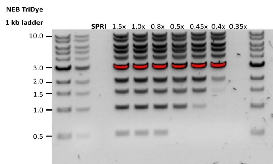 |
| Low recovery after end-prep | The wash step used ethanol <70% | DNA will be eluted from the beads when using ethanol <70%. Make sure to use the correct percentage. |
14. Issues during the sequencing run
Below is a list of the most commonly encountered issues, with some suggested causes and solutions.
We also have an FAQ section available on the Nanopore Community Support section.
If you have tried our suggested solutions and the issue still persists, please contact Technical Support via email (support@nanoporetech.com) or via LiveChat in the Nanopore Community.
Fewer pores at the start of sequencing than after Flow Cell Check
| Observation | Possible cause | Comments and actions |
|---|---|---|
| MinKNOW reported a lower number of pores at the start of sequencing than the number reported by the Flow Cell Check | An air bubble was introduced into the nanopore array | After the Flow Cell Check it is essential to remove any air bubbles near the priming port before priming the flow cell. If not removed, the air bubble can travel to the nanopore array and irreversibly damage the nanopores that have been exposed to air. The best practice to prevent this from happening is demonstrated in this video how to load a PromethION Flow Cell. |
| MinKNOW reported a lower number of pores at the start of sequencing than the number reported by the Flow Cell Check | The flow cell is not correctly inserted into the device | Stop the sequencing run, remove the flow cell from the sequencing device and insert it again, checking that the flow cell is firmly seated in the device and that it has reached the target temperature. If applicable, try a different position on the device. |
| MinKNOW reported a lower number of pores at the start of sequencing than the number reported by the Flow Cell Check | Contaminations in the library damaged or blocked the pores | The pore count during the Flow Cell Check is performed using the QC DNA molecules present in the flow cell storage buffer. At the start of sequencing, the library itself is used to estimate the number of active pores. Because of this, variability of about 10% in the number of pores is expected. A significantly lower pore count reported at the start of sequencing can be due to contaminants in the library that have damaged the membranes or blocked the pores. Alternative DNA extraction or purification methods may be needed to improve the purity of the input material. The effects of contaminants are shown in the Contaminants know-how piece. Please try an alternative extraction method that does not result in contaminant carryover. |
MinKNOW script failed
| Observation | Possible cause | Comments and actions |
|---|---|---|
| MinKNOW shows "Script failed" | Restart the computer and then restart MinKNOW. If the issue persists, please collect the MinKNOW log files and contact Technical Support. If you do not have another sequencing device available, we recommend storing the flow cell and the loaded library at 4°C and contact Technical Support for further storage guidance. |
Pore occupancy below 40%
| Observation | Possible cause | Comments and actions |
|---|---|---|
| Pore occupancy <40% | Not enough library was loaded on the flow cell | The minimum recommended library loaded here is ~100 ng as DNA is sheared. Check Qubit measurements and perform fragment length QC to troubleshoot. |
| Pore occupancy close to 0 | Ethanol was used instead of LFB or SFB at the wash step after sequencing adapter ligation | Ethanol can denature the motor protein on the sequencing adapters. Make sure the LFB or SFB buffer was used after ligation of sequencing adapters. |
| Pore occupancy close to 0 | No tether on the flow cell | Tethers are added during flow cell priming (FCT tube). Make sure FCT was added to FCF before priming. |
Shorter than expected read length
| Observation | Possible cause | Comments and actions |
|---|---|---|
| Shorter than expected read length | Unwanted fragmentation of DNA sample | Read length reflects input DNA fragment length. Input DNA can be fragmented during extraction and library prep. 1. Please review the extraction methods in the Nanopore Community for best practice for extraction. 2. Visualise the input DNA fragment length distribution on an agarose gel before proceeding to the library prep. 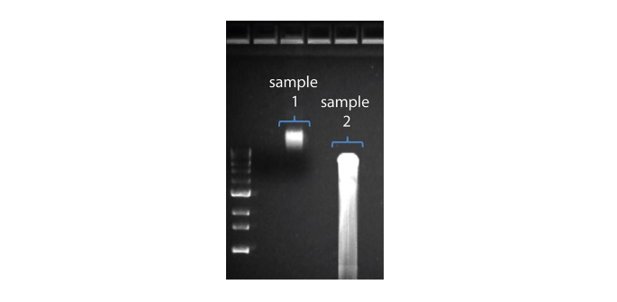 In the image above, Sample 1 is of high molecular weight, whereas Sample 2 has been fragmented. 3. During library prep, avoid pipetting and vortexing when mixing reagents. Flicking or inverting the tube is sufficient. |
Large proportion of unavailable pores
| Observation | Possible cause | Comments and actions |
|---|---|---|
Large proportion of unavailable pores (shown as blue in the channels panel and pore activity plot) 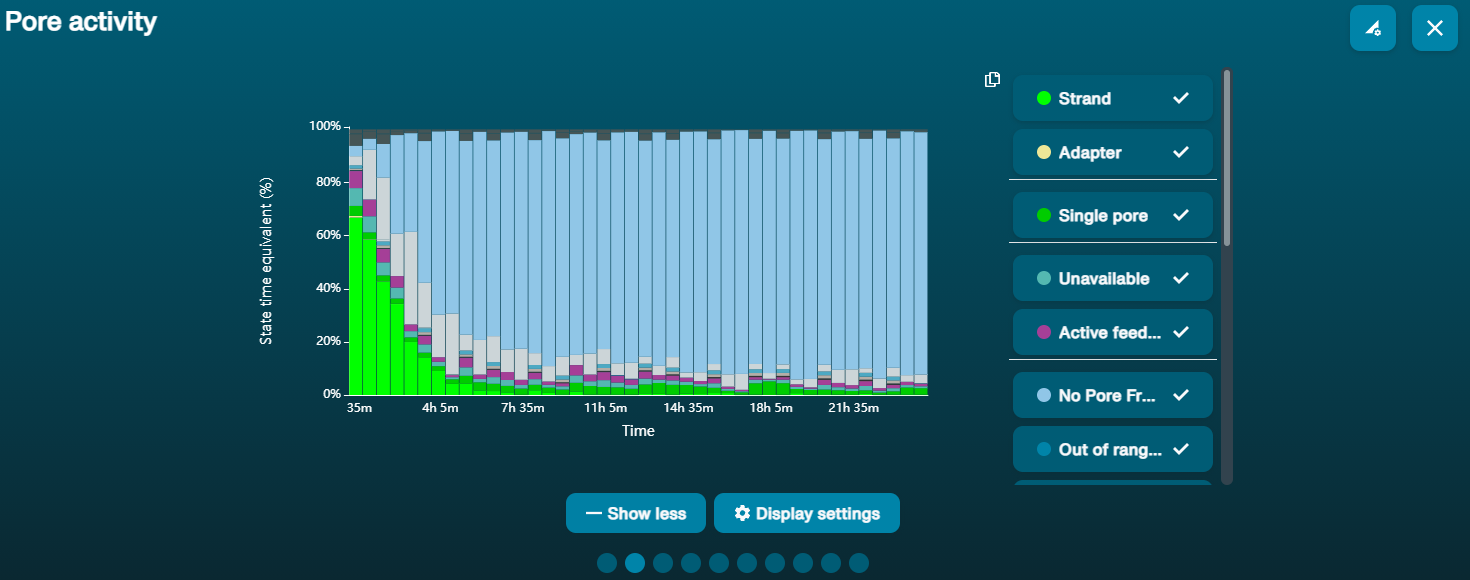 The pore activity plot above shows an increasing proportion of "unavailable" pores over time. | Contaminants are present in the sample | Some contaminants can be cleared from the pores by the unblocking function built into MinKNOW. If this is successful, the pore status will change to "sequencing pore". If the portion of unavailable pores stays large or increases: A nuclease flush using the Flow Cell Wash Kit (EXP-WSH004) can be performed. |
Large proportion of inactive pores
| Observation | Possible cause | Comments and actions |
|---|---|---|
| Large proportion of inactive/unavailable pores (shown as light blue in the channels panel and pore activity plot. Pores or membranes are irreversibly damaged) | Air bubbles have been introduced into the flow cell | Air bubbles introduced through flow cell priming and library loading can irreversibly damage the pores. Watch the how to load a PromethION Flow Cell video for best practice. |
| Large proportion of inactive/unavailable pores | Certain compounds co-purified with DNA | Known compounds, include polysaccharides. Clean-up using the QIAGEN PowerClean Pro kit. |
| Large proportion of inactive/unavailable pores | Contaminants are present in the sample | The effects of contaminants are shown in the Contaminants know-how piece. Please try an alternative extraction method that does not result in contaminant carryover. |
Temperature fluctuation
| Observation | Possible cause | Comments and actions |
|---|---|---|
| Temperature fluctuation | The flow cell has lost contact with the device | Check that there is a heat pad covering the metal plate on the back of the flow cell. Re-insert the flow cell and press it down to make sure the connector pins are firmly in contact with the device. If the problem persists, please contact Technical Services. |
Failed to reach target temperature
| Observation | Possible cause | Comments and actions |
|---|---|---|
| MinKNOW shows "Failed to reach target temperature" | The instrument was placed in a location that is colder than normal room temperature, or a location with poor ventilation (which leads to the flow cells overheating) | MinKNOW has a default timeframe for the flow cell to reach the target temperature. Once the timeframe is exceeded, an error message will appear and the sequencing experiment will continue. However, sequencing at an incorrect temperature may lead to a decrease in throughput and lower q-scores. Please adjust the location of the sequencing device to ensure that it is placed at room temperature with good ventilation, then re-start the process in MinKNOW. |
Last updated: 11/11/2026
ドキュメントオプション