Salmon (Salmo salar) tissue DNA

Extraction Method
Salmon (Salmo salar) tissue DNA
FOR RESEARCH USE ONLY.
Introduction
This is a method to extract and sequence high molecular weight genomic DNA from the Atlantic salmon (Salmo salar), as an example of fish tissue. We found tissue samples generated lower sequencing outputs compared to samples extracted from blood stored in ethanol at a final concentration of 90%. However, we observed that shearing samples using the Covaris g-TUBE TM or the Megaruptor® could also be used to increase sequencing output but at the expense of read N50. For further general data showing performance comparison of fragmented gDNA, please see ‘Optional Fragmentation of gDNA’. In order to obtain maximum sequencing data from the flow cell, we recommend using the Flow Cell Wash Kit. Sequencing performance was assessed using the Ligation Sequencing Kit.
Materials
- ~25 mg of brain, heart, liver, spleen, or fin tissue stored at −80°C prior to gDNA extraction
- MagAttract HMW DNA kit
- Magnetic rack
- ThermoMixer™
- Vortex
- 1.5 ml and 2 ml Eppendorf tubes
- Microcentrifuge
- TE buffer (10 mM Tris-HCL, 1 mM EDTA, pH 8.0)
- Ice and ice bucket
- Weighing boats
- Tweezers and scalpel
Method
Use up to 25 mg of tissue to cut into small pieces and grind up using tweezers and a scalpel. The tissues should be allowed to thaw, but we recommend this process should be carried out as quickly as possible with the weighing boat placed on ice to grind the tissue sample.
Transfer the tissue to a 2 ml Eppendorf tube containing 220 µl of ATL buffer.
Add 20 µl of proteinase K and vortex to mix.
Incubate on the ThermoMixer™ at 56°C for 3 hours at 900 rpm to agitate. The tissue should be completely lysed at this point. However, if there are tissue chunks present in the solution, centrifuge the tube at 20,000 x g for 3 minutes and transfer the supernatant to a fresh Eppendorf tube to take forward. We recommend avoiding carrying through any tissue that has not been lysed.
Add 4 µl of RNase A and flick the tube to mix. Incubate at room temperature for 3 minutes.
Follow the recommended protocol from step 3 to step 13 of the handbook (pages 24-25).
Add 150 µl of TE and incubate the sample at room temperature for 30 minutes.
Incubate the sample on the ThermoMixer™ at 37°C for 1 minute at 1400 rpm.
Briefly centrifuge the tube and place it on the magnetic rack.
Transfer the supernatant containing the DNA to a fresh 1.5 ml Eppendorf tube.
Repeat steps 7-11 twice more, using fresh Eppendorf tubes each time.
Results
Table 1. gDNA quantification and QC data. Genomic DNA extracted from various salmon tissues was quantified using the Qubit fluorometer and purity was assessed using the NanoDrop.
| Sample | Yield (µg) | A260/280 | A260/230 |
|---|---|---|---|
| Blood | 6-16 | 1.92 | 0.43 |
| Heart | 25-35 | 1.79 | 1.91 |
| Liver | 70-80 | 2.00 | 2.25 |
| Spleen | 60-70 | 1.81 | 2.00 |
| Fin | 50-60 | 1.95 | 2.37 |
| Brain | 30-40 | 1.55 | 1.02 |
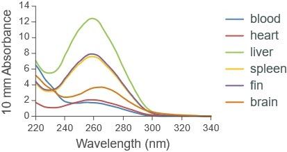
Figure 1. NanoDrop spectra for gDNA extracted from various salmon tissues
Sequencing performance
Libraries were prepared using the Ligation Sequencing Kit.
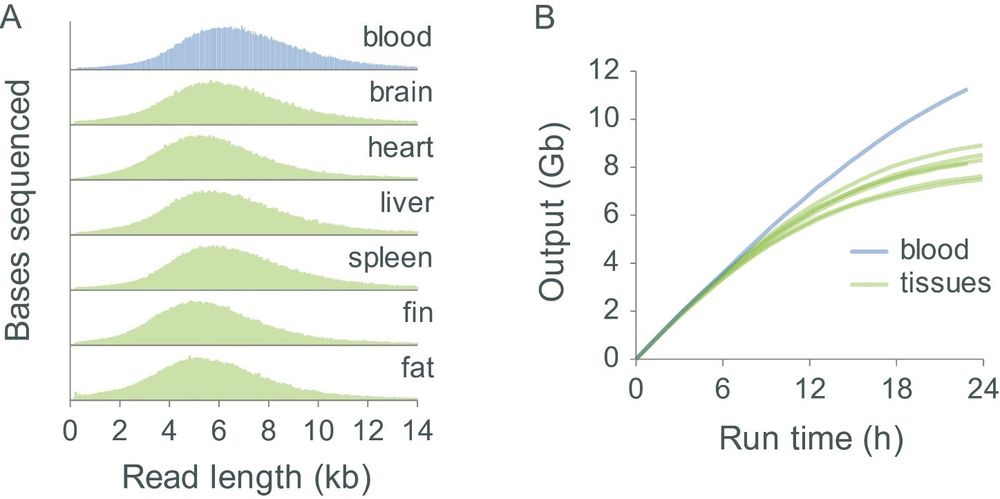
Figure 2. Sequencing performance of gDNA extracted from various salmon tissues. Extracted gDNA was sheared using the Covaris g-TUBE to normalise fragment lengths between the various tissues and libraries were prepared using the Ligation Sequencing Kit and sequenced on MinION for 24 hours. Panel A: The read length distributions obtained from the blood and other tissue samples shows that read lengths were successfully normalised by the shearing procedure. Panel B: Higher sequencing outputs were obtained using gDNA extracted from blood rather than gDNA extracted from tissue samples.
Change log
| Version | Change |
|---|---|
| v1, 28th July 2021 | Initial protocol publication |


