Buffy coat DNA

Extraction Method
Buffy coat DNA
FOR RESEARCH USE ONLY.
Contents
Introduction
Materials
Method
Results
Sequencing performance
Change log
Introduction
This protocol describes a method to extract high molecular weight genomic DNA from buffy coat, which has been isolated from rabbit blood, as an example of mammalian blood. The buffy coat is the fraction of an anticoagulated blood that contains white blood cells and platelets following centrifugation. Sequencing performance was determined by a MinION™ using the Ligation Sequencing Kit. Prior to library preparation, 3 µg of extracted DNA was size selected using SPRI beads.
Materials
- 10 ml rabbit blood (Note: The buffy coat should be isolated, as soon as possible after blood collection)
- 15 and 50 ml Falcon tubes
- 1x phosphate buffer saline (PBS)
- Ficoll®-Paque PREMIUM
- Centrifuge (with adjustable deceleration) with capacity for 15 and 50 ml Falcon tubes
- QIAGEN MagAttract HMW DNA kit
- Eppendorf ThermoMixer
- 1.5 and 2 ml Eppendorf tubes
- Magnetic rack
- Vortex
- TE (10 mM Tris-HCl, 1 mM EDTA, pH 8.0)
Method
- If the blood has been stored after collection (e.g. at 4°C, -20°C or -80°C), allow it to equilibrate at room temperature for ~2 hours. Note: The buffy coat should be isolated via the process below, as soon as possible after blood collection.
- Add 10 ml of blood to a 50 ml Falcon tube.
- Add 10 ml of 1x PBS and mix gently by inverting 5 times.
- Add 12 ml of Ficoll to the bottom of a fresh 50 ml Falcon tube.
- Gently overlay the blood (Step 3) onto the Ficoll (Step 4), without breaking Ficoll’s surface.
- Centrifuge for 20 minutes at 760 x g, at room temperature. The deceleration must be set to zero. Note: It is important that the deceleration is set to zero to prevent mixing of the plasma and interface layers.
- Remove as much of the plasma, i.e. the top layer, as soon as possible to reveal the interface containing the white cells. Collect the white cells by pipette aspiration.
- Transfer the white cells to a fresh 15 ml Falcon tube.
- Top up to 15 ml with (1x) PBS.
- Centrifuge for 8 minutes at 350 x g, at room temperature. Note: It is no longer important that the deceleration is set to zero.
- Carefully remove and discard the supernatant. Retain the pellet.
- Repeat steps 9-11 two more times.
- At this point, the protocol may be paused, and the cell pellet may be frozen at -80°C.
- With wide-bore tips, pipette and transfer the pellet to a 2 ml Eppendorf tube and add 40 µl of proteinase K
- Add 8 µl of RNase A and 300 µl of buffer AL to the pellet solution, and vortex to homogenise.
- Incubate the homogenised pellet at room temperature for 30 minutes.
- Add 30 µl of MagAttract Suspension G and 360 µl of Buffer MB to the lysate.
- Place the tube in the ThermoMixer for shaking and incubation at room temperature for 3 minutes at 1400 rpm.
- Follow Steps 8a to 15 according to the QIAGEN MagAttract HMW DNA kit manual (pages 16-17). We recommend that the elution is performed with 200 µl of TE.
a. Place the tube holder of the MagAttract Magnetic Rack on its magnetic base, wait until bead separation has been completed (~1 minute), and remove the supernatant.
b. Add 700 µl Buffer MW1 to the sample and place the tube holder of the MagAttract Magnetic Rack onto the mixer. Incubate at room temperature for 1 minute at 1400 RPM. To increase the efficiency of the wash step, remove the tube holder from magnetic base before adding the wash buffer. Add the wash buffer directly onto the magnetic bead pellet.
c. Place the tube holder of the MagAttract Magnetic Rack on its magnetic base, wait until bead separation has been completed (~1 minute), and remove the supernatant.
d. Repeat steps b. and c.
e. Add 700 µl Buffer PE to the sample and place the tube holder of the MagAttract Magnetic Rack onto the mixer. Incubate at room temperature for 1 minute at 1400 RPM. To increase the efficiency of the wash step, remove the tube holder from the magnetic base before adding the wash buffer. Add the wash buffer directly onto the magnetic bead pellet.
f. Place the tube holder of the MagAttract Magnetic Rack on its magnetic base, wait until bead separation has been completed (~1 minute), and remove the supernatant.
g. Repeat steps e. and f.
h. Rinse the particles with 700 µl distilled water while the tube holder is on the magnetic base and the beads are fixed to the wall of the sample tube. Incubate for 1 minute at room temperature, and remove the supernatant completely. Do not pipette water directly onto the bead pellet. Pipette it into the sample tube against the side facing away from the bead pellet. All pipetting steps must be performed carefully to avoid disturbing the fixed bead pellet.
i. Repeat step h.
j. Remove the tube holder of the MagAttract Magnetic Rack from its magnetic base and add 200 µl of TE buffer. Place the tube holder onto the mixer and incubate at room temperature for 3 minutes at 1400 RPM.
- Take 3 µg of eluate and perform a SPRI size selection.
Results
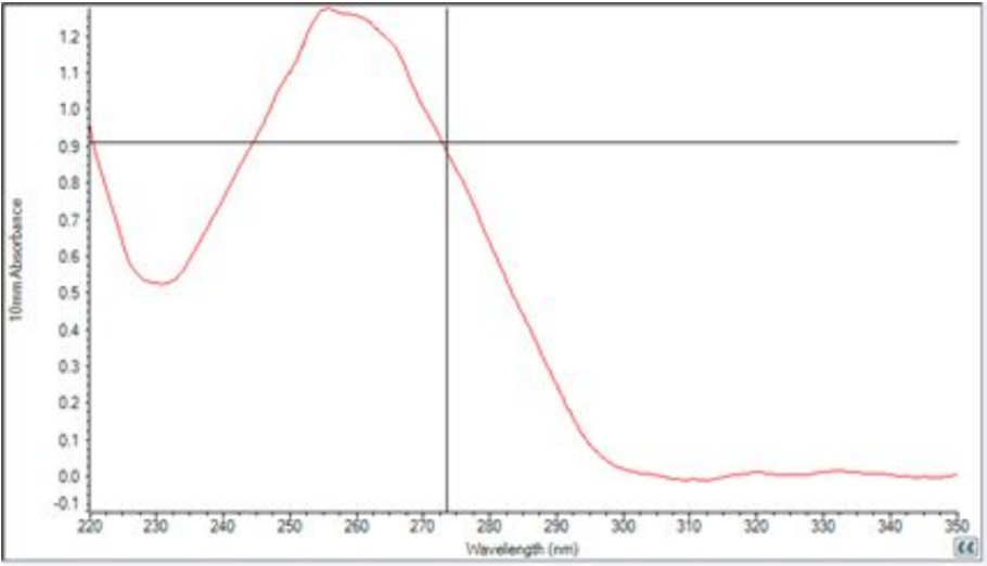
- Yield: 4-6 μg
- OD 260/280: 1.97
- OD 260/230: 2.41
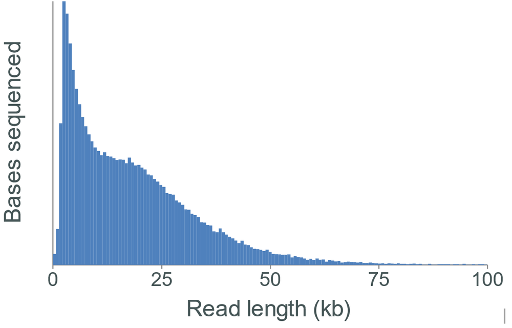
Sequencing performance
Libraries were prepared from unsheared and sheared DNA using the Ligation Sequencing Kit.
Change log
| Version | Change |
|---|---|
| v1, 26th June 2019 | Initial protocol publication |
| v2, 14th August 2023 | Updated recommended steps to follow in the QIAGEN handbook and wrote out the steps in full. |


